Download
1 / 63
640 likes | 675 Views
Tissue Repair: Regeneration, Healing and Fibrosis. Dr.Ahmad abualsamen. TISSUE REPAIR Restoration of tissue architecture & function after an injury. It occurs by two reactions: Regeneration Healing by fibrosis. 1- Regeneration:. Replacement of the diseased tissue

E N D
Tissue Repair: Regeneration, Healing and Fibrosis Dr.Ahmad abualsamen
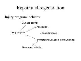
TISSUE REPAIR • Restoration of tissue architecture & function after an injury. • It occurs by two reactions: • Regeneration • Healing by fibrosis.
1- Regeneration: Replacement of the diseased tissue By proliferation of parenchymal cells. Results in complete restoration.
II- Repair by laying down of connective (fibrous) tissue: It occurs if the injured tissues are: Incapable of proliferation The supporting structures of the tissue are severely damaged.
organization Fibrosis in a tissue space occupied by inflammatory exudate E.g. organizing pneumonia & serofibrinous inflammation in pleura.
The Cell Cycle • Rate of cell division: different • Some cells do not divide. • Other cells cycle every 16-24 hours. • Mitosis is controlled by genes • Genes encode the release of specific proteins • This protiens promote or inhibit mitosis at different steps.
Mitosis-promoting proteins: • Cyclins. • Cyclins activate cyclin-dependent kinases (CDKs) • CDK acts in conjunction with cyclins • regulate the phosphorylation of proteins involved in cell cycle progression • leading to DNA replication and mitosis. • After mitosis is completed, cyclins & CDKs are degraded
Cell cycle is tightly regulated by stimulators and inhibitors, Contains intrinsic checkpoint controls to prevent replication of abnormal cells. Interphase : Period between two mitoses
According to the proliferative capacity of the cells in tissue Tissues are divided into: Labile Stable Permanent
Continuously dividing tissues (labile tissues) contain: Stem cells that differentiate to Replenish lost cells Maintain tissue homeostasis.
Stem Cells Unspecialized cells Can divide through mitosis Differentiate into specialized cell types. Self-renew to produce more stem cells.
Types of stem cells: Embryonic stem (ES) cells: Isolated from the inner cell mass of blastocyst Differentiate into ALL cells of the adult Preserve small populations of more restricted stem cells Adult stem cells; Found in various tissues Differentiation into ALL CELL TYPES OF THEIR TISSUEi.e: Possess a more restricted range of cell differentiation than ES cells).
Factors affecting Cell proliferation tissue repair List the factors that influence wound healing A- Many chemical mediators, such as growth factors, hormones, and cytokines B- Interactions between cells and ECM components
Growth factors(Table 4.1) Is a protein that has the following effects; Stimulate cellular PROLIFERATION. Stimulate Migration, Differentiation & Contractility. Enhance the synthesis of Specialized Proteins (such as collagen in fibroblasts). Stimulate the function of growth control Genes (Proto-oncogenes).
Mechanisms of action. Induce cell proliferation by binding to specific receptors. Affect the expression of genes ===products promote replication. Enhance the synthesis of cellular proteins involved in mitosis. Prevent apoptosis
Extracellular matrix (ECM) • Dynamic component • Synthesized locally • Assembled into network surrounds cells. Synthesis and degradation of ECM accompanies : • Morphogenesis. • Wound healing. Chronic fibrotic processes. Tumor invasion & metastasis.
ECM serves several important fuctions: Mechanical support to tissues Substrate for cell growth and the formation of tissue Microenvironments. Regulates cell proliferation &differentiation. Intact ECM is required for tissue regeneration, if ECM is damaged, repair can only be accomplished by scar formation.
Forms of ECM Interstitial Matrix: This is present in the spaces between cells in connective tissue, and between epithelium and supportive vascular and smooth muscle structures. It is synthesized by mesenchymal cells (e.g., fibroblasts). Its major constituents are fibrillar and nonfibrillar collagens, as well as fibronectin, elastin, proteoglycans, hyaluronate, and other elements. Basement Membrane The basement membrane lies beneath the epithelium and is synthesized by overlying epithelium and underlying mesenchymal cells. Its major constituents are amorphous nonfibrillar type IV collagen and laminin.
Main Types of Collagens Tissue Distribution, typeII : Cartilage, intervertebral disk • TypeIII : Soft tissues • Type V : Soft tissue and blood vessels • Type1V : Basement Membrane • Type1X : Cartilage
Components of the ECM There are three basic components of ECM: 1- Fibrous structural proteins: such as collagens and elastins, give tensile strength and recoil. 2- Water-hydrated gels such as proteoglycans & hyaluronan, permit flexibility and lubrication. 3- Adhesive glycoproteins: Connect matrix elements to one another & to cells.
Role of the ECM Provides mechanical support to tissues (role of collagen &elastin). Control of cell growth & proliferation by signaling through cellular receptors of the integrin family. Maintenance of cell differentiation. The type of ECM proteins can affect the degree of differentiation of the cells via cell surface integrins. Scaffolding for tissue renewal; the integrity of the basement membrane or the stroma is critical for regeneration of tissues, whose disruption leads to scar formation. Establishment of tissue microenvironments; basement membrane acts as a boundary between epithelium and underlying connective tissue and also forms part of the filtration apparatus in the kidney. Storage & presentation of regulatory molecules e.g. growth factors fibroblast GF & hepatocyte GF are excreted and stored in the ECM. This allows for the rapid use of growth factors after local injury, or during regeneration.
Extensive regeneration can occur only if • Residual tissue is structurally & functionally intact • If the tissue is damaged by inflammation, regeneration is incomplete and is accompanied by scarring.
Repair by connective tissue: This occurs if: Non-dividing (permanent) cells are injured. Tissue injury is severe or chronic, Damage to parenchymal cells and epithelium as well as the stromal framework.
Cells involved Mesenchymal cells (C.T. Stem cells), Endothelial cells Macrophages, platelets Parenchymal cells of the injured organ.
Repair by connective tissue deposition consists of four sequential processes: • Angiogenesis • Migration and proliferation of fibroblasts. • Deposition of ECM (scar formation). • Maturation & reorganization of the fibrous tissue (remodeling).
Cutaneous wound healing Involves: Epithelial regeneration Connective tissue scar. Based on the nature of the wound, thehealing can occur by : First Second intention.
Healing by first intention (Primary union) Healing of a wound which: Clean and uninfected. Surgically incised. Without much loss of cells & tissue. Edges of wound are approximated by surgical sutures
The sequence of events in primary union Initial haemorrhage: Immediately after injury Space is filled with blood Blood clots seals the wound against dehydration &infection.
Within 24 hours; • Neutrophils at the incision margin & toward the fibrin clot. • Basal cells at the cut edge of the epidermis begins mitosis.
Within 24 to 48 hours; Epithelial cells migrate & proliferate along the dermis Deposition of basement membrane components as they progress. The cells meet in the midline as a thin but continuous epithelial layer separating the underlying viable dermis from the overlying scab.
By day 3: Granulation tissue invades the incision space. Collagen fibers formation , Vertically oriented Do not bridge the incision. Epithelial cell proliferation continues
Morphology of granulation tissue Grossly: granulation tissue appears Granular Soft & Moist Pink and bleeds on touch. Insensitive Resistant to bacterial infection
Microscopy; it is characterized by: Proliferation of fibroblasts New thin-walled delicate capillaries (angiogenesis) In a loose ECM.
By day 5: Granulation tissue fills the incisional space Neovascularization reaches its peak Collagen fibrils : abundant & bridge the incision. Epidermisrecovers its normal thickness: Mature epidermal architecture with surface keratinization (skin).
During the second week: Fibroblast proliferation & collagen accumulation. Leucocytic infiltrate, edema & vascularity diminished. By the end of the first month:Scar
Initial haemorrhage: Blood clots seals wound Within 24 hours; Neutrophils, Basal cells 24 to 48 hours; Epithelial cells proliferate , deposition of basement membrane , thin but continuous epithelial layer separating dermis from the scab.. By day 3: Granulation tissue,Collagen fibers, Epithelial cell proliferation By day 5: Granulation tissue fills space, Collagen fibrils , Epidermisnormal thickness. During the second week: collagen, inflammation By the end of the first month:Scar
By the end of the first month: Scar Comprises acellular connective tissue Devoid of inflammatory cells Covered by a normal epidermis. Dermal appendages destroyed in the line of the incision are permanently lost. Tensile strength of the wound increases with time
In second-intention healing, the basic events are similar to primary union but differ in: Defect : larger . Hence healing takes place from the base upwards & from the margins inwards. Inflammatory reaction more intense Granulation tissue abundant Followed by accumulation of ECM Formation of a large scar Wound contracts within 6 weeks May be reduced to 5 -10% of its original size (due to the presence of myofibroblasts (modified fibroblasts).
Wound Strength • Carefully sutured wounds have approx. 70% of the strength of normal skin. • When sutures are removed, usually at one week, wound strength is approximately 10% of that of normal skin, but this increases rapidly over the next 4 weeks. • Recovery of tensile strength results from collagen synthesis exceeding degradation during the first 2 months, and from structural modifications of collagen (e.g., cross- linking and increased fiber size). • Wound strength reaches approximately 70% -80% of normal by 3 months but usually does not improve beyond that point.
Factors affecting wound healing: Infection: most important cause of delayed wound healing. Foreign bodies such as fragments of steel, glass, or even bone impair healing. Nutrition : protein & vitamin C deficiency inhibit collagen synthesis & delay healing.
Poor perfusion, due to arteriosclerosis, diabetes or obstructed venous drainage results in impaired healing. • Glucocorticoids (steroids) • Have anti-inflammatory effects, and their administration may result in poor wound strength due to diminished fibrosis. • In some cases , e.g. corneal infections, glucocorticoids are sometimes prescribed (along with antibiotics) to reduce the likelihood of opacity that may result from collagen deposition.