Download
1 / 50
500 likes | 650 Views
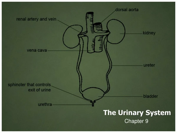
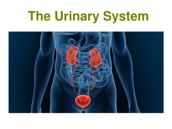
The Urinary System. Chapter 18. 8 31 2012 online ed. Urinary System. Also called “excretory system” Consists of: Two kidneys Two ureters One urinary bladder One urethra. Kidneys. 2 bean shaped bodies situated behind peritoneum

E N D
The Urinary System Chapter 18 8 31 2012 online ed.
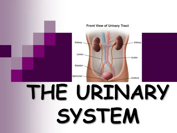
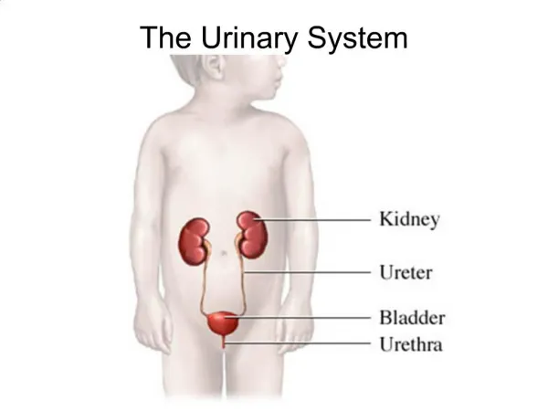
Urinary System Also called “excretory system” Consists of: • Two kidneys • Two ureters • One urinary bladder • One urethra
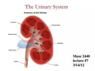
Kidneys 2 bean shaped bodies situated behind peritoneum Asymmetrical - left is slightly longer and narrower than right Why is Rt kidney slightly lower than Lt kidney? Liver Both lie in an oblique plane (opposite sijt direction) Normally extend from T-12 to L3
Kidney Function • Remove waste products from blood • Maintain fluid and electrolyte balance • Secrete substances that affect blood pressure • How much urine excreted per day? 1 - 2 liters
Kidneys (cont’d) • Minor calyces unite to form major calyces • Major calyces unite to form renal pelvis • Renal pelvis drains into ureters • Hilum - longitudinal slit in medial border for transmission of blood vessels, nerves, lymphatic vessels, and ureter
Kidneys (cont’d) Each kidney has: cortex medulla Medulla contains collecting system Essential microscopic components of kidney called nephrons Each kidney-contains how many nephrons? about 1 million
Anatomy: Nephron • Glomerulus - filter for blood, allows fine particles and water to pass into capsule • Renal tubule is continuous with capsule • Proximal convoluted tubule • Nephron loop (loop of Henle) • Distal convoluted tubule • Distal convoluted tubule opens into collecting ducts • Collecting ducts drain into minor calyx
Adrenal Glands(Suprarenal) Not part of urinary system Cannot be seen on plainradiographs (need CT) Regulate stress response through release of various hormones such as adrenaline
Ureters • Two tubes 10 - 12 “ long • Retroperitoneal • Extend from renal pelvis • Enter bladder at ureteral orifice • How is urine moved through ureters? • peristalsis
Urinary Bladder • Musculomembranous sac situated immediately posterior and superior to symphysis pubis of pelvis • Serves as Urine reservoir
How much fluid can bladder hold? • up to 500 mL • Internal rethral orifice located in bladder neck • Area between ureteral openings and urethral orifices is trigone
Urethra • Carries urine from bladder to? • exterior of body • How long is it in females? • About 1.5 • In males? • About 7 to 8 • Sphincter at neck of bladder • Male urethra contains following parts: • Prostate • Membranous area • Spongy area
IVU- Intravenous Urogram ! Formerly erroneously known as IVP-Intravenous pyelogram! pyelo refers to renal pelvis and calyces only But study also shows ureters, bladder, and sometimes urethra
Indications For Urography • Demonstrate physiologic function and structure of urinary system • Evaluate abd. Masses, renal cysts and tumors • Urolithiasis (stones) • Pyelonephritis (Inflammation of kidney) • Hydronephrosis (distension of renal pelvis and calyces with urine) • Trauma • Renal hypertension • Pre-op evaluation
Contraindications • Inability to filter contrast medium from blood • Allergy to contrast • Abnormal BUN and Creatinine levels
Preparation Of Pt • Pt on low residue diet for 1-2 days prior to exam • Laxative taken day prior to clean out bowel • NPO after midnight • Pts with multiple myeloma, high uric acid levels, or diabetes should be well hydrated before IVP exam (Dehydration leads to increased risk of renal failure)
Contrast Media • Used to visualize urinary tract adequately • Iodinated, water-soluble contrast administered intravenously • Antegrade filling
Contrast Media Excretory urography (IVU) generally uses a 50 to 70% iodine solution Lower concentrations required for bladder studies due to large amount required to fill bladder (30%) Non-ionic contrast is generally used More expensive, but- Patients less likely to have reactions with nonionic
Contrast Media and Adverse Reactions Do not leave pt. alone for first 5 minutes after injection! Mild reactions: warmth flushing hives, Nausea/Vomiting, respiratory edema (accumulation of fluid in lungs) Severe reactions: Anaphylactic shock: sudden allergic response: sudden drop in blood pressure and difficulty breathing Death in a matter of minutes
IVU Procedure Scout – KUB Contrast injected Timed sequence of films obtained until bladder begins to fill Take Immediate image of kidneys 5 minute image of abd. or kidneys Then apply Compression (Take tomograms)
Ureteral Compression • (Because of improvement of contrast agents, compression no longer generally used) • Compression device centered at ASIS over distal ends of ureters With as much compression as pt can tolerate! • Inhibits flow of urine into bladder • Distends renal pelvis and calyces
Contraindications for Compression Should not be applied when pt has: Kidney stones abdominal mass or aneurysm colostomy suprapubic catheter recent abd. surgery or trauma
Radiation Protection Gonadal shield - if it does not interfere with exam Shield males for all urinary studies, except when urethra is of primary interest Shield females when IR centered over kidneys Close collimation Avoid repeat exposures Rule out chance of pregnancy before examination (Emergency cases may not allow time)
AP Projection-IVU • Patient supine • Typical Abdomen positioning • Use shielding • (All exposures at end of expiration for any urinary system study)
AP Projection- IVU (cont’d) Must include entire KUB region Should include prostatic region on older males
Time Delay - IVU 3 minute 6 minutes
Time delay- IVU With compression 9 minutes
AP Projection Variations Trendelenberg: Lower head 15 - 20 degrees Helps demonstrate lower ureters Upright: Must lower CR - organs change position Prone Demonstrates ureteropelvic region Fills obstructed ureter in cases of hydronephrosis(distension of renal pelvis and calyces with urine)
AP Oblique Projections - RPO/LPO • Pt. supine Rotated 30 degrees • Typical Abdomen oblique position
AP Oblique Projections (cont’d) • Elevated kidney will be parallel to cassette • Kidney closest to cassette will be perpendicular • Entire KUB region must be included
Nephrotomography • Best method for visualizing renal parenchyma (neprons and collecting tubules) • To visualize kidneys free of intestinal content superimposition
Tomogram Procedure cont’d • Tomograms are obtained once bladder is filled • Pt is measured, divide number by 3, cuts begin there • Pt. measures 30cm, beginning cuts at 10cm • Release compression slowly • Have pt void, and obtain post-void film
Retrograde Urography What does retrograde mean? Requires catheterization of ureters Contrast injected directly through cathethers Provides improved opacification of renal collecting system Opposite normalflow
Retrograde Urography (cont’d) • Contrast does not enter blood stream • Used for patients with renal insufficiency or contrast sensitivity • Ureters, and collecting systems can be selectively imaged and sampled • Little physiologic information provided
Retrograde Urography cont’d • Considered an operative procedure • Pt may be under general anesthesia • Sterile technique • Nurse responsible for set-up of exam and pt. care
Cystography • Radiologic exam of urinary bladder • Contrast administration usually performed retrograde(against normal flow of urine)
Indications for Cystography Vesicoureteral reflux (backward flow of urine into ureters) Recurrent lower urinary tract infection Neurogenic bladder: (dysfunction due to disease of central nervous system or peripheral nerves) Bladder trauma Prostate enlargement Lower urinary tract fistulae Urethral stricture Posterior urethral valves (obstructive congenital defect of the male urethra)
Contraindications for Cystography Anything related to catheterization of urethra!
Cystography Procedure • Contrast drip-infused via a catheter • Bladder filled to capacity • Fluoro-spot and overhead images obtained
Routine Cystography Series Scout Filled AP or PA (axial) Both obliques Lateral Post-void
AP Axial Bladder(similar to coccyx projection) CR: Angle 10 to 15 degrees caudad Enters 2 above upper border of pubic symphysis Can be done PA
AP Oblique Bladder Pt position: 40- to 60-deg. rotation RPO or LPO depending on physician preference
Lateral Bladder Demonstrates: anterior/posterior bladder walls • Base of bladder • Any vesicovaginal or vesicorectal fistulae
Male Cystourethrography • Images obtained as contrast injected by urethral syringe • Entire urethra must be visualized • Bladder can be filled to obtain antegrade voiding study • Why is this antegrade if its injected into urethra? AP Oblique Projection - RPO/LPO
Female Cystourethrography • Retrograde • AP Projection (maybe obliques) • Bladder can be filled and pt. voids for antegrade studies
Voiding Cystourethrogram X-ray images of bladder and urethra during urination Follows cystogram - urinary catheter removed Pt. urinates into special radiolucent urinal as images taken .
Voiding Cystourethrogram cont’d • Shows size and shape of bladder under stress caused by urination • Demonstrates urethra functioning • Most commonly used for young girls with history of recurrent bladder infections
Metallic Bead Chain Cystourethrography • To evaluate stress incontinence in females only • Beaded chain inserted in Urethra • Shows anatomic changes in shape and position of anatomic floor • Valsalva tech. applied for comparison
Summation of exams of Urinary System IVU- entire urinary system Retrograde Urogram- same as IVU but performed through catheter starting at urethra Nephrotomography- slices of kidneys Cystogram- for bladder Voiding Cytogram Cystourethrogram- for urethra Voiding Cystourethrogram