Download
1 / 40
400 likes | 676 Views
Esophageal Cancer Rates (Men, Age Standardised Mortality/105). USA. . 20100. . US SEER data, 2004. . Esophageal Cancer Rates (Men, Age Standardised Mortality/105). . LondonUKUSA. . 20100. . Thames Cancer Registries

E N D
1. Dr Laurence Lovat
National Medical Laser Centre
University College London, UK
2. Esophageal Cancer Rates (Men, Age Standardised Mortality/105)
3. Esophageal Cancer Rates (Men, Age Standardised Mortality/105)
4. Esophageal Cancer Rates (Men, Age Standardised Mortality/105)
5. The Questions How do we prevent death from esophageal adenocarcinoma?
How do we detect patients at risk?
How do we best treat patients?
6. The Questions How do we prevent death from esophageal adenocarcinoma?
How do we detect patients at risk?
How do we best treat patients?
7. Detecting patients at risk 80% of cases of esophageal adenocarcinoma arise within Barrett�s esophagus
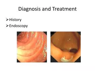
Screening
Endoscopy
Invasive
Not of proven value
Less invasive techniques
Nothing proven
8. Detecting patients at risk Surveillance
Lifetime risk of cancer <10%
Need to target high risk groups
9. Markers of Risk
10. Cancer Risk in Presence of Aneuploidy or Tetraploidy >6%
11. p16, p53, ploidy Biomarker Panel Progression to EA
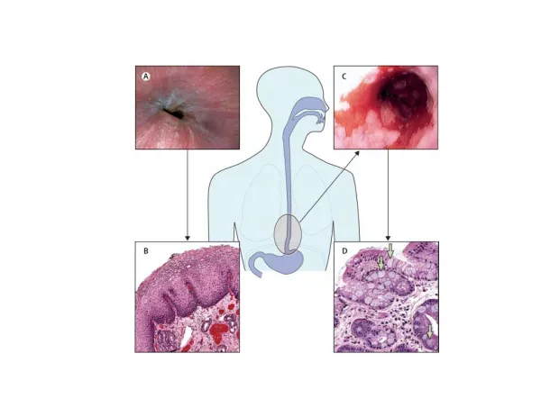
12. Endoscopic Detection of HGD
13. Morphological Changes in HGD
14. Elastic Scattering Spectroscopy(�Optical biopsy�) Point measurement
Wavelength dependence
Scattering efficiency of tissue
Sensitive to morphological changes
Size, shape and density of nuclei & mitochondria
cellular density
15. Elastic Scattering Spectroscopy
16. Optical Biopsy
17. Elastic Scattering Spectra Separate evidence in vitrofrom rat fibroblasts that amound of genetic material in cells
affects the slope between 630 and 820 nmSeparate evidence in vitrofrom rat fibroblasts that amound of genetic material in cells
affects the slope between 630 and 820 nm
19. ESS �Optical Biopsies� Perform 17 random biopsies
OR
17 optical measurements and 7 biopsies
Model:
92% dysplasia sites detected
Negative test >99.5% reliable
20. Taking Optical Biopsy Forward Detecting patients at high risk who do NOT have dysplasia
Can OB detect patients at risk
21. Detecting �Field Change� Effect Animal model of colon cancer
Aberrant crypt foci is the first visible change
ESS detects �fingerprint� of microarchitectural abnormalities BEFORE aberrant crypt foci visible
( Roy et al, Gastroenterology (2004); 126: 1071)
Human Colorectal Cancer Risk Stratification
37 patients
Colonoscopy in those with/without previous adenomas
Similar findings to animal models
( Roy et al, DDW 2005)
22. The Questions How do we prevent death from esophageal adenocarcinoma?
How do we detect patients at risk?
How do we best treat patients?
23. Treatment for HGD in Barrett�s Oesophagus Oesophagectomy
Morbidity � 40% Mortality � 5%
Elderly patients
Need for minimally invasive therapy
Aim
Show the problems of conventional treatment
Quote number not fit for surgery
Standard treatment of
total oesophagectomy
High morbidity and significant mortality
High risk to the patient of prolonged stay and of death
Its still needed, but �Hugh Barr�s quote
Need for minimally invasive treatment
QUOTE on another slide? With figure of oesophagectomyAim
Show the problems of conventional treatment
Quote number not fit for surgery
Standard treatment of
total oesophagectomy
High morbidity and significant mortality
High risk to the patient of prolonged stay and of death
Its still needed, but �Hugh Barr�s quote
Need for minimally invasive treatment
QUOTE on another slide? With figure of oesophagectomy
24. Mucosal Ablation Thermal (hot/cold)
Laser
MPEC
Cryotherapy
Photochemical (PDT)
25. The Ideal of Mucosal Ablation
Selective mucosal
destruction
Ambulatory therapy
No side effects Strictures Photosensitivity Acute Hypotension Buried glands
28. PDT Results: Barrett�s Esophagus
29. Results (ALA) From October 1999
75 patients treated
(most after 2002)
All had high grade dysplasia (V4)
3 studies:
High dose ALA (60mg/kg)
Light dose ranging (low, medium, high light dose)
RCT ALA 30 mg/kg with red v green light
RCT ALA 60 mg/kg with red v green light UpdateUpdate
30. ALA 60 mg/kg (high dose):Red Light at various doses
31. ALA 30 mg/kg (low dose):Red v Green Light
32. Rescue with high dose ALAand various light doses
33. ALA PDT 75 patients treated
Best regime: 80% clearance HGD at 2 years
Toxicity (all at 60mg/kg)
4 patients: severe hypotension
(prevented by rehydration and avoiding psychotropic drugs)
3 patients: aspiration pneumonia
8 patients: transient fever
2 patients: asymptomatic jaundice, cleared in 5 days
34. ALA PDT Looks promising but there are toxicity issues
35. Foscan Mucosal Selectivity Explain the box-plot
Interquartile range
MedianExplain the box-plot
Interquartile range
Median
36. Verteporfin photosensitiser(2mg/kg, activated at 15 minutes) Ivc injection, full bladder!Ivc injection, full bladder!
37. Duodenal PDT Histology 10 J to pancreas (therefore adjacent damage). Slide on left shows loop of duodenum with complete necrosis of duodenal mucosa. Slides on right ( low and high power)10 J to pancreas (therefore adjacent damage). Slide on left shows loop of duodenum with complete necrosis of duodenal mucosa. Slides on right ( low and high power)
38. Duodenal collagen resistant to damage
39. The Ideal of Mucosal Ablation
Selective mucosal
destruction
Ambulatory therapy
No side effects Strictures Photosensitivity Acute Hypotension Buried glands
40. Conclusions Optical methods might be developed to detect patients at highest risk
New PDT approaches to treat HGD in BE
Can optical methods be used to assess the outcome of PDT?