Download
1 / 47
1.22k likes | 2.42k Views
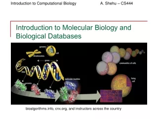
Introduction to molecular biology. Genes, proteins and DNA. A nineteenth-century monk called Gregor Mendel introduced the notion of genes : basic units responsible for possession and passing on of a single characteristic Initially it was thought that proteins carried genetic information

E N D
Genes, proteins and DNA • A nineteenth-century monk called Gregor Mendel introduced the notion of genes: basic units responsible for possession and passing on of a single characteristic • Initially it was thought that proteins carried genetic information • Until mid 20th-century, when it was found that DNA did. • Proteins are the functional molecules in cells (ie. they perform the majority of the reactions of life)
What is DNA? • Built up of a sugar-phosphate backbone and a sequence of nucleotides: adenine (A), guanine (G), cytosine (C) and thymine (T) • Watson and Crick discovered the structure of DNA: a double helix– two sugar phosphate chains wrapping round each other, with the nucleotides sticking out – the nucleotide from strand 1 meets the nucleotide from strand 2 in the middle. These pairs of nucleotides are complementary – where one strand has a C, the other has a G and vice versa; where one strand has an A the other has a T and vice versa. • Human DNA consists of approximately 3 x 10⁹ such “base pairs”.
DNA replication • The DNA molecule is directional, because the sugars are asymmetrical – each sugar is connected to the strand “upstream” at its 5th carbon and “downstream” at its 3rd carbon. So you read the DNA sequence from the “5 prime” end to the “3’ ” end. • In replication, the double helix becomes unzipped and free nucleotides bind to the their complementary pair nucleotides on the single strands. Thus each strand acts as a template for a new strand of DNA. The reaction is catalysed by DNA polymerase- this causes the chain to elongate, but it can’t start the formation of a new chain. For this a primer(short piece of DNA/ RNA) is required.
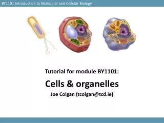
What is a cell? • Structural unit of most organisms • Chemical factory enclosed by a semi-permeable membrane • Different types of cell within an animal • Prokaryotic cells are simple structurally • Likely precursors of more complex eukaryotic cells • Eg. Bacteria • No compartments within the cell such as nucleus- just cytosol with plasma membrane • Eukaryotic cells are compartmentalised- they contain organelles which perform specific functions – eg. nucleus, mitochondria.
Organelles • The nucleus – houses the chromosomal DNA which is the genetic information store • The rough endoplasmic reticulum– where most of theribosomesreside, which are the sites for protein synthesis • Mitochrondria – are the powerhouses of the cell, producing energy for the reactions of life • See http://www.biology.arizona.edu/cell_bio/tutorials/pev/page3.html for more details on organelles.
Chromosomes • In eukaryotes, the DNA is stored in the nucleus. Since there is not much space, the DNA must be highly organised. • The DNA double helix is wrapped around protein complexes called histones- each unit of DNA wrapped round a histone is called a nucleosome. • To condense the DNA further, nucleosomes are grouped together to form chromatin fibres. • The chromatin fibres fold together to form large looped domains • These looped domains are then organised into distinct structures called chromosomes. • Many eukaryotic cells contain pairs of chromosomes and are hence called diploid. • Other cells contain single chromosomes and are called haploid. • The human genome consists of 23 pairs of chromosomes.
The central dogma • DNARNAprotein TranscriptionTranslation • The expression of genetic information stored in DNA involves, first transcription into RNA and then translation into the functional protein molecules, in which the amino acid sequence is determined by the nucleotide sequence of the DNA.
Genes • A gene is a region of DNA that controls a discrete hereditary characteristic, usually corresponding to a single mRNA which will be translated into a protein. • In eukaryotes, the genes have their coding sequences (exons) interrupted by non-coding sequences (introns). • In humans, genes constitute only about 2-3% of DNA, the rest is “junk” DNA.
RNA • RNA is like DNA but the sugar-phosphate backbone has a different sugar: ribose instead of deoxyribose • and where the DNA molecule has the nucleotide thymine (T), RNA has the nucleotide uracil (U). • RNA is almost always a single stranded molecule whereas DNA always stored as a double helix in eukaryotes. • RNA comes in different forms including: • Messenger RNA (mRNA) is transcribed from DNA and translated into protein • Transfer RNA (tRNA) is a functional molecule used in the process of translation (see later)
Transcription • The process of production of RNA from DNA is called transcription; it consists of three stages: • Initiation – the RNA polymerase enzyme binds to a promoter site on the DNA and unzips the double helix. • Elongation – free nucleotides bind to their complementary pairs on the template strand of the DNA elongating the RNA chain which is identical to the informational strand of DNA, except that the nucleotide thymine in DNA is replaced by uracil in RNA. The polymerase moves along the DNA in the 3’ to 5’ direction, extending the RNA 5’ to 3’. • Termination – specific sequences in the DNA signal termination of transcription; when one of these is encountered by the polymerase, the RNA transcript is released from the DNA and the double helix can zip up again.
Splicing The original transcript from the DNA is called heavy nuclear RNA (hnRNA). It contains transcripts of both introns and exons. The introns are removed by a process called splicing to produce messenger RNA(mRNA) and the ends of the RNA molecule are processed.
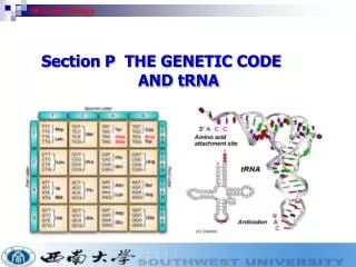
Translation • Special molecules called transfer RNAs (tRNAs) recognise both an amino acid and a triplet of nucleotides (a codon). The tRNA molecule has an anticodon on one end which binds to a codon on the mRNA and to a specific amino acid on the other end. It thus enforces the genetic code in which a codon codes for a specific amino acid. • Protein synthesis takes place on the ribosomes. The tRNAs position themselves for reading the genetic message in the mRNA. The first tRNA binds to a start codon (AUG) on the mRNA and then each tRNA adds an amino acid to a growing polypeptide (protein) chain.
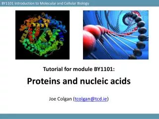
What is a protein? • A protein is a linear polymer of amino acids linked together by peptide bonds. • The average protein is c. 200 amino acids long, but some can contain thousands of amino acids. • Proteins are the main functional chemicals in the cell, carrying out many functions, for example catalysis of the reactions involved in metabolism. • Proteins have a complex structure which can be thought of as having four structural levels.
Protein structure • Primary structure – the sequence of amino acids in the protein chain • Secondary structure – the local spatial arrangement of the protein; short stretches of the chain fold up to form structures such as alpha-helices and beta-sheets • Tertiary structure – the long range 3D structure of the chain – how the beta-sheets, etc. relate to each other in space ( they pack into domains) • Quaternary structure – a protein may consist of more than one linear chain molecule; the quaternary structure determines how these chains fold around one another
The amino acids Alanine Ala A Cysteine Cys C Aspartic AciD Asp D Glutamic Acid Glu E Phenylalanine Phe F Glycine Gly G Histidine His H Isoleucine Ile I Lysine Lys K Leucine Leu L Methionine Met M AsparagiNe Asn N Proline Pro P Glutamine Gln Q ARginine Arg R Serine Ser S Threonine Thr T Valine Val VTryptophan Trp W Tyrosine Tyr Y Proteins are polymers of the 20 naturally occurring amino acids. Each amino acid has a three-letter code and a single letter code:
The amino acids • Consist of a central carbon atom (the alpha-carbon) connected to an amino group, a carboxyl group, a hydrogen atom and a side chain. The side chain differs between the different amino acids but the rest is the same: • Have different properties because their side chains have different shapes and chemical groups. • Hydrophobic/hydrophilic, acidic/basic/neutral, aliphaptic/aromatic, conformationally important http://web.mit.edu/esgbio/www/lm/proteins/aa/aminoacids.html
Example amino acids Grey=carbon, white=hydrogen, red=oxygen, blue=nitrogen Proline: C5H9NO2 Side chain: C3H6 , links to N in amino group Alanine: C3H7NO2 Side chain: CH3 http://www.cryst.bbk.ac.uk/PPS2/course/section2/index.html , Sami Raza
The peptide bond AminoAcid1 + AminoAcid2 Dipeptide + Water , ie. • Polypeptides are just long chains of amino acids linked by peptide bonds. Proteins are made up of one or more polypeptide chains (cf. quaternary structure). • Name comes from peptide group –CONH-. H | (AminoAcid1) - C=O + H-N+ - (AminoAcid2) (AminoAcid1) - C=O + H2O | | | O- H H-N-(AminoAcid2) peptide bond
Primary structure • Primary structure of a protein is simply the linear sequence of amino acid in its polypeptide chain(s) (NB proteins are written in order from the amino-teminal end to the carboxy-terminal end, so Ala-Cys-Phe is different from Phe-Cys-Ala) • Eg. first 100 (of 457) amino acids in hexokinase: A A S X D X S L V E V H X X V F I V P P X I L Q A V V S I A T T R X D D X D S A A A S I P M V P G W V L K Q V X G S Q A G S F L A I V M G G G D L E V I L I X L A G Y Q E S S I X A S R S L A A S M X T …… • This sequence contains all the information required to determine the higher levels of structure. The linear polypeptide chain folds in a particular arrangement, giving a three-dimensional structure, but the information on how to fold is contained in the sequence.
Alpha helix • An alpha-helix is a tight rodlike helix formed out of the polypeptide chain. The polypeptide main chain makes up the central structure, and the side chains extend out and away from the helix. The CO group of one amino acid (n) is hydrogen bonded to the NH group of the amino acid four residues away (n +4):
Alpha helix • Alpha helices are most commonly made up of hydrophobic amino acids, because hydrogen bonds are generally the strongest attraction possible between such amino acids. • Between one amino acid and the next is a rise of 1.5Å and a turn of 100. • Alpha helices are found in almost all proteins to various extents , eg. haemoglobin -75% (see Stryer). • Proteins have right-handed helices
Beta Sheet • A beta pleated sheet is another type of secondary structure. Sheets can either be parallel or anti-parallel. Anti-parallel sheet looks like this: With hairpin turns like this (hydrogen bonds between amino acid n and n+4):
Beta sheet • The individual lines of amino acids within the protein are called strands. • Typically a sheet will have 2-5 parallel or anti-parallel strands. • The axial distance between two amino acids is 3.5Å.
Loops • Between the alpha helices and beta strands in the protein are loop regions. • These are less regular, although may still have some structure. • Loops tend to end up on the outside of the proteins when the protein folds up to form its full 3d structure (tertiary structure), so they are exposed to water- the loop regions thus tend to be rich in hydrophilic residues (amino acid side chains)- cf. prediction. • Loops often ~ binding sites for other molecules.
Schematic diagrams of secondary structure • Secondary structure elements are often represented in ribbon diagrams: Coiled ribbon= alpha helix, arrow ribbon= beta strand, thin string= loops, etc. VHL protein Stebbins et al, Science, 284:455.
A protein domain • The tertiary structure is the way in which the secondary structure elements (eg. alpha helices) fit together in the full 3d structure. • The protein folds up so that amino acids which are far apart in the linear sequence may be close together in space, forming a domain: http://www.cryst.bbk.ac.uk/PPS2/course/section10/1pfk.gif
Disulphide bridges • The tertiary structure also describes the pattern of disulphide bridges, which form between the 2 cysteine (C) amino acids. Cysteine (like methionine) contains a sulphur atom and when two cysteines are spatially close they can form covalent –S-S- bonds: http://www.chem.uwec.edu/Chem406/Webpages/Ying/overview.htm
A protein with more than one chain • Insulin is also a good example of a protein with more than one polypeptide chain: • In fact the structure of insulin is more complicated: it forms hexamers with six insulin molecules (A and B chain) around two zinc ions and 6 water molecules. In the quaternary structure, the B chain is wrapped around the A chain, which forms a compact central unit.
Mutation • Replication of DNA is pretty accurate, but not error-free. Occasionally mutations take place: ATCGGGCCATATCGAAATGG ATCGCGCCATTTCGAAATGG • In humans, mutation rate may be c. 3 mutations per genome (3 billion bps) per cell division. • Actually this error is what allows evolution to take place, since it leads to the generation of novel genotypes (cf. genetic algorithms). mutations
Recombination • Recombination is the crossing over of DNA sequence between two copies of a chromosome. It occurs in the egg and sperm so that each chromosome you receive from your mother(/father) has a mixture of her(/his) mother’s and her(/his) father’s DNA: Chromosome 1 ACTGA|CCCTATCGG Chromosome 2 TTGGC|AGCTAGCTG Offspring 1 ACTGA|AGCTAGCTG Offspring 2 TTGGC|CCCTATCGG
Recombinant DNA technology- gene cloning • Plasmids ~ small circular pieces of DNA found in bacteria. They are separate from bacterial chromosome. • Restriction enzymes can be used to cut the two strands of the plasmid at particular places leaving loose ends. • The DNA of interest, eg. human DNA for producing insulin protein can then be cut out of its chromosome (short linker sequences can be added to the ends to make a perfect match) and then stuck into the gap in the plasmid ring • The recombinant DNA formed is then put back in the bacteria where it is replicated • When the bacteria divide their offspring share the recombinant plasmids and produce many copies: in this way many copies of the foreign gene are cloned.
Gene expression • All cells in your body have the same genomic DNA (up to a very small mutational error), ie. the sequences of nucleotides within the chromosomes are identical. • How then do different cells maintain very different characteristics? • The answer is that not all of the genes in the genome are being transcribed and translated into proteins in every cell. We say that genes which are transcribed & translated are expressed in the cells. • Gene expression controls distinct identities of cells via functional protein molecules (cf. microarrays).
Conclusions • This has been a general introduction to molecular biology, introducing the key molecules of life: • DNA (the store of genetic information) • RNA • & protein (the function molecules of the cell) • Central Dogma: DNA is transcribed to form RNA which is translated to form protein • Key processes: DNA replication, transcription, translation
Conclusions • We discussed DNA and protein structure (which are important for their functions). • We introduced: • Concepts of mutation and recombination, which are fundamental to evolution and will be important when we discuss sequence comparsions • Recombinant DNA technology: important in medicine and for our next topic- sequencing • Gene expression: gives cells their distinct identities and important for block 3 (microarrays) [see http://www.chemie.fu-berlin.de/chemistry/bio/amino-acids.html for formulae for all the amino acids]