Download
1 / 30

E N D
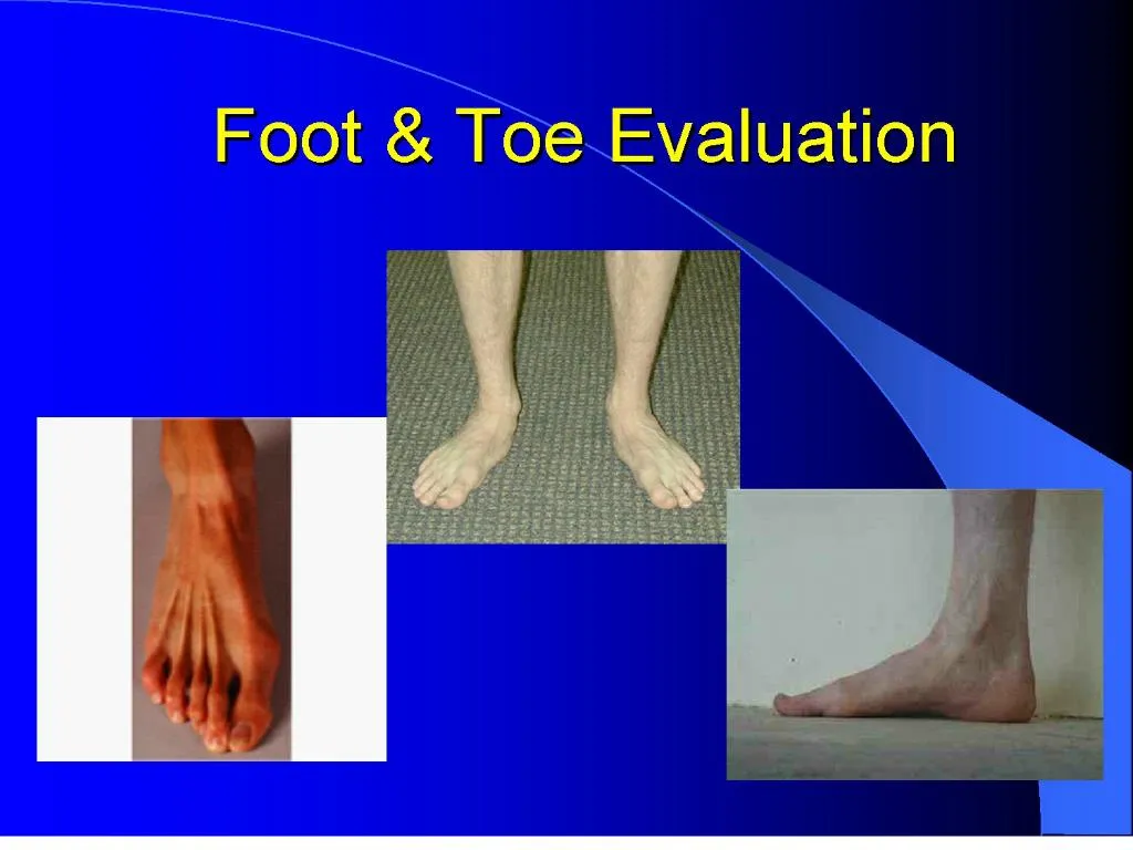
1. Foot & Toe Evaluation
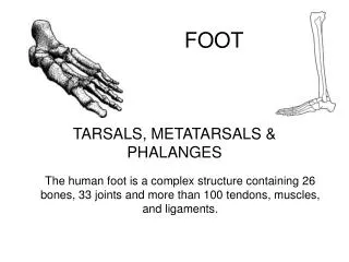
2. Foot Anatomy 26 Bones
Rearfoot � Calcaneus, Talus
Midfoot � 3 Cuneiforms, Cuboid, Navicular
Forefoot � 5 Metatarsals, 14 Phalanges, 2 Sesamoids
3. Ankle mortise � tibia, fibula, talus Reference: Starkey, C. (2002). Evaluation of Orthopedic & Athletic Injuries, p. 89.
4. Rearfoot Provides stability & shock absorption during initial stance phase
Lever arm for Achilles tendon during plantarflexion
Talus � no muscles attach to it
Calcaneus
- Calcaneal tubercle � posterior side
- Sustentaculum tali �
medial side - helps support the talus
flexor hallicus longus - passes through the medial
groove
- Peroneal tubercle � lateral side
Subtalar joint � inferior talus/superior calcaneus
5. Rearfoot Reference: Primal Pictures, 2001
6. Midfoot Shock absorber
Medial longitudinal arch
Navicular tuberosity � Tibialis posterior insertion
7. Forefoot Lever during pre-swing phase
Metatarsals � proximal base, body, distal head
Phalanges � MTP, PIP, DIP, IP joints
Plantar fascia
8. Forefoot
9. Metatarsophalangeal Joints Classified as condyloid-type joints
Great toe metatarsophalangeal (MP) joint flexes 45� & extends 70�
MP joints of the four lesser toes
40� of flexion
40� of extension
also abduct & adduct minimally
10. Joints Great toe interphalangeal (IP) joint flexes from 0� of full extension to 90� of flexion
Proximal interphalangeal (PIP) joints in lesser toes flexes from 0� extension to 35� flexion
Distal interphalangeal (DIP) joints flexes 60� & extend 30�
Much variation from joint to joint & from person to person
11. Terminology Intrinsic muscles � m. contained within the foot
Extrinsic muscles � m. originating from lower leg or femur
Supination � inversion + adduction + plantarflexion
Pronation - eversion + abduction + dorsiflexion
Inversion � movement of the plantar aspect of the calcaneus toward the midline of the body
Eversion � movement of the plantar aspect of the calcaneus away from the body
Dorsiflexion � flexion of the ankle, pulling the foot & toes toward the tibia
Plantarflexion � extension of the ankle, pointing the foot & toes
Ray � series of bones formed by the MT & phalanges
12. Movements of Foot Pronation
combination of ankle dorsiflexion, subtalar eversion, & forefoot abduction (toe-out)
Supination
combination of ankle plantar flexion, subtalar inversion, & forefoot adduction (toe-in)
Eversion
turning ankle & foot outward; abduction, away from midline; weight is on medial edge of foot
Inversion
turning ankle & foot inward; adduction, toward midline; weight is on lateral edge of foot
13. Movements of Toes Toe flexion
movement of toes toward plantar surface of foot
Toe extension
movement of toes away from plantar surface of foot
14. Intrinsic Muscles of the Foot All originate & insert within the foot
Extensor digitorum brevis - dorsum of foot
Remainder are in a plantar compartment in 4 layers on plantar surface of foot
15. Intrinsic Muscles of the Foot First (superficial) layer: Abductor hallucis, flexor digitorum brevis, abductor digiti minimi (quinti)
Second (middle) layer: Quadratus plantae, lumbricales (4)
16. Intrinsic Muscles of the Foot Third (deep) layer: Flexor hallucis brevis, adductor hallucis, flexor digiti minimi brevis
Fourth (deep) layer: Dorsal interossei (4), plantar interossei (3)
17. Intrinsic Muscles of the Foot Grouped by location
Medial - attach to great toe proximal phalanx
Abductor hallucis & flexor hallucis brevis - medially
Adductor hallucis - centrally beneath metatarsals
Central location
Beneath the foot
Quadratus plantae, 4 lumbricales, 4 dorsal interossei, 3 plantar interossei, flexor digitorum brevis
Dorsal compartment
Extensor digitorum brevis
Lateral � attach on lateral aspect of base of 5th phalange proximal phalanx
abductor digiti minimi, flexor digiti minimi brevis
18. Intrinsic Muscles of the Foot Grouped by action
4 muscles act on great toe
abductor hallucis - abduction of great toe & assists flexor hallucis brevis in flexing great toe at MP joint
adductor hallucis - adduction of great toe
extensor digitorum brevis - extension of great toe at MP joint
4 lumbricales
flexors of the 2nd, 3rd, 4th, & 5th phalanges at MP joints
quadratus plantae
flexors of 2nd, 3rd, 4th, & 5th phalanges at DIP joints
3 plantar interossei
adductors & flexors of proximal phalanxes of 3rd, 4th, & 5th phalanges
4 dorsal interossei
abductors & flexors of 2nd, 3rd, & 4th phalanges MP joints
flexor digitorum brevis
flexes middle phalanxes of 2nd, 3rd, 4th, & 5th phalanges
extensor digitorum brevis
extends great toe & 2nd, 3rd, 4th phalanges at MP joints
5th toe muscles
abductor digiti minimi abducts proximal phalanx
flexor digiti minimi brevis flexes proximal phalanx
19. Lateral Foot & Ankle
20. Medial Foot & Ankle
21. Extrinsic Muscles of the Foot Extensor hallucis longus (EHL)
Extensor digitorum longus (EDL)
Flexor hallucis longus (FHL)
Flexor digitorum longus (FDL)
Triceps Surae (Gastrocnemius, Soleus, Plantaris)
Peroneus longus, brevis, tertius
Tibialis anterior
Tibialis posterior
Starkey, Ch. 4, Table 4-2, p. 92-95
22. Neurovascular Nerves
Tibial n. � medial side
Lateral & Medial Cutaneous branch (comes from Peroneal n.) � lateral side
Vascular
Tibial a.
Dorsalis Pedis pulse
23. Lateral Cutaneous Distribution Peach � Sural N.
Purple � Lat. Plantar N.
Yellow � Superficial Peroneal N.
24. Medial Cutaneous Distribution Light Green � Saphenous N.
Yellow � Superficial Peroneal N.
Dark Pink � Tibial N. (Medial Calcaneal branches)
Dark Green � Medial Plantar N.
25. Dermatome Distribution Green � L4
Pink � L5
Salmon � S1
26. Arches Ligaments in foot & ankle maintain arches
Two longitudinal arches
Medial longitudinal arch - extends from calcaneus bone to talus, navicular, 3 cuneiforms, and proximal ends of 3 medial metatarsals
Lateral longitudinal arch - extends from calcaneus to cuboid and proximal ends of 4th & 5th metatarsals
Transverse arch
extends across foot from 1st metatarsal to the 5th metatarsal
27. Evaluation of the Foot History �
What happened? (MOI)
Where is the pain?
When did it happen? (onset)
Has it happened before?
What does it feel like?
Pain scale (1-10)
What type of surface?
How old are the shoes?
Type of pain
Unusual noises/sensations Observation �
Toes, Arches
Forefoot & Rearfoot Valgus/Varus, Pronation/Supination
Calluses, blisters, warts, etc.
Appearance
Bilateral comparison
Color
Deformity
Edema, Swelling
Gait
Infection
Weight bearing vs. non-weight bearing
Shoe wear pattern
28. Evaluation of the Foot Palpation �
Start away from the point of pain
Palpate bony & soft tissue structures
Medial structures
Lateral structures
Dorsal structures
Plantar structures
Crepitus
Heat
Swelling
Rigidity
Deformities
Softness Stress Tests �
ROM tests (AROM, PROM, RROM-strength)
Alignment
Ligament & Capsular tests
Fracture tests
Neurological tests
Other special tests
29. Assessment & Plan What injury have you evaluated?
What are you going to do with this injury?
30. Common Injuries Retrocalcaneal bursitis
Heel contusion
Arch strains
Plantar fasciitis
Fractures
Bunion
Know the signs & symptoms of these injuries Sesamoiditis
Morton�s neuroma
Sprained toes � Turf toe
Fractures & dislocations