Download
1 / 36
510 likes | 1.51k Views
Patent Ductus Arteriosus. By Dr. Hanan Zekri Khaled Lecturer of pediatrics Pediatric cardiology. Patent Ductus Arteriosus. Patent Ductus Arteriosus. Patent Ductus Arteriosus.

E N D
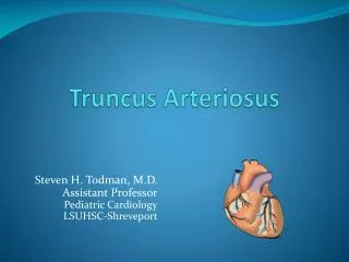
Patent Ductus Arteriosus By Dr. Hanan Zekri Khaled Lecturer of pediatrics Pediatric cardiology
Patent Ductus Arteriosus ●The ductus arteriosus in the fetus is an important conduit that allows deoxygenated blood to bypass the collapsed lungs and enter the placenta through the descending aorta and umbilical arteries.
Patent Ductus Arteriosus ● The placenta acts as an oxygenator and returns oxygen rich blood through the umbilical vein and ductus venosus to the fetal heart.
Patent Ductus Arteriosus ● The placenta produces prostaglandins, which maintain prenatal patency of the ductus and, in early gestation, inhibit the ability of the ductus to contract in response to oxygen.
Patent Ductus Arteriosus ● The ductus arteriosus itself also produces prostaglandins and nitric oxide-like vasodilators.
Patent Ductus Arteriosus ● During the postnatal period, final closure of the ductus arteriosus results from increased production of local vasoconstrictors (like endothelin) in response to higher arterial oxygen, removal of placental prostaglandin and a decrease in the number of prostaglandin E2 receptors in the ductal wall.
Patent Ductus Arteriosus • During the first 60 hours of life, spontaneous closure of the ductus occurs in 55% of full-term newborn infants. • By 2-6 months of age, closure occurs in more than 95% of healthy infants.
Patent Ductus Arteriosus • Persistent patency of the ductus arteriosus following birth is inversely related to gestational age. • This may be due to the smaller amount of muscular tissue in the media with lower intrinsic tone, and lower responsiveness to oxygen but higher sensitivity to the vasodilating effects of prostaglandin E2 and nitric oxide.
Reopening of a Constricted Ductus • Before true anatomic closure occurs, the functionally closed ductus may be dilated by a reduced arterial Po2 or an increased PGE2 concentration. The reopening of the constricted ductus may occur in asphyxia and various pulmonary diseases (as hypoxia and acidosis relax ductal tissues).
Patent Ductus Arteriosus • The direction of blood flow across the PDA depends on the balance of pulmonary and systemic vascular resistance.
Patent Ductus Arteriosus • The most reliable non-invasive diagnostic tool is echocardiography with Doppler ultrasound. • In most infants, a modified parasternal short axis view offers the best window for PDA visualization. • This view offers the best opportunity to directly measure the PDA.
Patent Ductus Arteriosus • The secondary effects of the increased flow can estimate the volume load from the left to right ductal shunt. • A large shunt leads to dilation of the left atrium and left ventricle, as well as holodiastolic reversal of blood flow distal to the ductus in the descending aorta due to run off into the pulmonary bed.
Patent Ductus Arteriosus • Enlargement of the left atrium reflecting the approximate magnitude of the shunt, can be further supported by left atrial-to-aortic root ratio of >1.3. • In addition, continuous wave Doppler can estimate pulmonary artery pressures by measuring Doppler velocities of PDA flow and tricuspid regurgitation.
Patent Ductus Arteriosus • The clinical features depend on the magnitude of left-to-right shunt through the PDA and the ability of the infant to initiate compensatory mechanisms to handle the extra volume load.
Patent Ductus Arteriosus • Because many premature infants have respiratory distress syndrome, the stage of development of this disease and the use of surfactant replacement therapy will determine the pulmonary vascular resistance and therefore the shunt.
Patent Ductus Arteriosus • The maturity of the infant and the stage of myocardial development determine the ability to handle the shunt.
Relationship to Systemic Organ Perfusion with PDA • Redistribution of systemic blood flow occurs even with moderate shunts. • Retrograde aortic flow, decreased systemic blood flow, and moderate hypotension are common in premature infants with a PDA and may lead to decreased perfusion in many organs, with potential clinical consequences to each.
Relationship to Systemic Organ Perfusion with PDA • Reduced cerebral blood flow or changes in cerebral blood flow velocity patterns have been implicated in the occurrence of intraventricular hemorrhage. • Renal function may be compromised, and myocardial perfusion, particularly subendocardial blood flow, may be reduced.
Treatment of PDA • In the premature infant an important aspect of PDA management is fluid intake. Early fluid restriction to allow for little more than insensible and sensible losses will significantly reduce the risks of PDA, necrotizing enterocolitis, and death at the expense of postnatal weight loss.
Treatment of PDA • Simple fluid restriction along with diuretic use is often recommended to control the symptoms of a PDA. • Furosemide is commonly used. Although furosemide is a prostaglandin agonist, it does not interfere with PDA closure.
Treatment of PDA • Furosemide merely helps the lungs clear fluid and thereby improves the patient’s ability to tolerate the PDA. Short-term use of furosemide and fluid restriction requires a close vigilance to prevent dehydration. Mehta SK, Younoszai A, Pietz J, Achanti BP. Pharmacological closure of the patent ductus arteriosus.Images Paediatr Cardiol 2003;14:1-15
Treatment of PDA • The use of oral or, preferably, intravenous (lyophilized) indomethacin to constrict the ductus arteriosus has led to successful nonsurgical closure in a large proportion of treated infants; the effects of indomethacin apparently are best when it is administered before 10 days of age and in less mature infants.
Treatment of PDA • Dose schedules vary, but commonly a first dose of 0.2 mg/kg is given by nasogastric tube or intravenously. • For intravenous indomethacin, subsequent doses depend on the age at initial treatment if <48 hours, the subsequent two doses are 0.10 mg/kg; if 2 to 7 days, 0.20 mg/kg; and if >7 days, 0.25 mg/kg. A total of three doses usually is given 12 to 24 hours apart depending on urinary output; if urine flow decreases, fewer doses may be used or the time between doses may be extended.
Treatment of PDA • If clinical signs reappear after an initially successful course of therapy, a second course may be considered.
Treatment of PDA • More recently, ibuprofen has also been evaluated as a possible alternative to indomethacin in preterm infants. • In addition, meta-analysis of the available studies has shown a comparable rate of ductal closure after ibuprofen treatment .
Treatment of PDA • Some evidence exists that there may be less effect of ibuprofen on renal function and urine output . • In addition, ibuprofen has less effect on cerebral vasculature and cerebral blood flow but has not shown a decreased risk for intraventricular hemorrhage.
Treatment of PDA Surgical ligation • In small infants that are not a candidate for, or who have failed, medical therapy, surgical ligation remains an effective alternative.
Treatment of PDA Trans-catheter closure • Although coil occlusion has been performed in infants, a large short PDA, which is the typical anatomy in symptomatic newborns and premature infants, is difficult to close.
Treatment of PDA • In addition, there is a significant risk of obstructing the descending aorta or left pulmonary artery, which are small caliber vessels in neonates.
Treatment of PDA • Out of the neonatal period, cardiac catheterization with coil occlusion of the PDA has become the primary mode of closure. • Newer occlusion devices similar to ones used for ASD closure are being developed for closure of the large PDA in older children and adults.
Treatment of PDA • Closure of a PDA by coil catheterization. (A) Injection into the aorta reveals a large PDA at baseline. (B) Following placement of a coil the angiographic dye no longer crosses into the pulmonary artery confirming ductal closure. (MPA = main pulmonary artery, PDA = patent ductus arteriosus, DA = descending aorta)