Download
1 / 1
10 likes | 178 Views
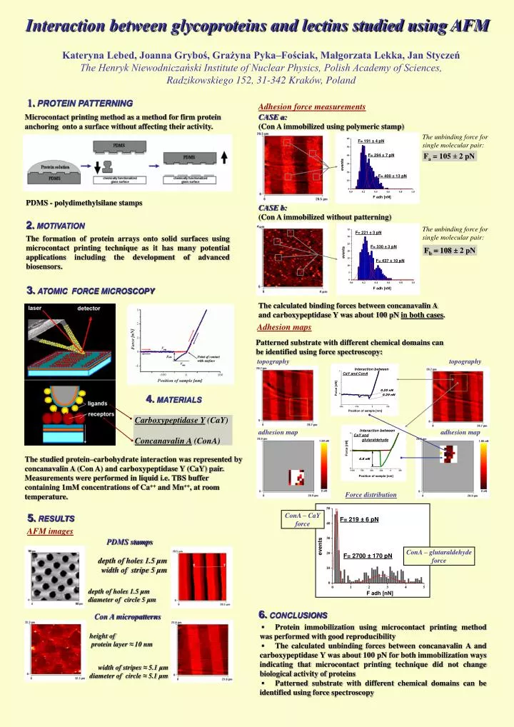
Interaction between glycoproteins and lectins studied using AFM. Kateryna Lebed, Joanna Gryboś, Grażyna Pyka–Fościak, Małgorzata Lekka, Jan Styczeń The Henryk Niewodniczański Institute of Nuclear Physics, Polish Academy of Sciences, Radzikowskiego 152, 31-342 Kraków, Poland.

E N D
Interaction between glycoproteins and lectins studied using AFM Kateryna Lebed, Joanna Gryboś, Grażyna Pyka–Fościak, Małgorzata Lekka, Jan Styczeń The Henryk Niewodniczański Institute of Nuclear Physics, Polish Academy of Sciences, Radzikowskiego 152, 31-342 Kraków, Poland 1.PROTEIN PATTERNING Adhesion force measurements Microcontact printing method as a method for firm protein anchoring onto a surface without affecting their activity. CASE a: (Con A immobilized using polymeric stamp) The unbinding force for single molecular pair: Fa = 105± 2 pN PDMS - polydimethylsilane stamps CASE b: (Con A immobilized without patterning) 2.MOTIVATION The formation of protein arrays onto solid surfaces using microcontact printing technique as it has many potential applications including the development of advanced biosensors. The unbinding force for single molecular pair: Fb= 108± 2 pN 3.ATOMIC FORCE MICROSCOPY The calculated binding forces between concanavalin A and carboxypeptidase Y was about 100 pN in both cases. Adhesion maps Patterned substrate with different chemical domains can be identified using force spectroscopy: topography topography 4.MATERIALS Carboxypeptidase Y (CaY)Concanavalin A (ConA) adhesion map adhesion map The studied protein–carbohydrate interaction was represented by concanavalin A (Con A) and carboxypeptidase Y (CaY) pair. Measurements were performed in liquid i.e. TBS buffer containing 1mM concentrations of Ca++ and Mn++, at room temperature. Forcedistribution 5.RESULTS ConA – CaY force AFM images PDMS stamps ConA – glutaraldehyde force depth of holes 1.5 µm width of stripe 5 µm depth of holes 1.5 µm diameter of circle 5 µm 6.CONCLUSIONS Con A micropatterns ▪ Protein immobilization using microcontact printing method was performed with good reproducibility ▪ The calculated unbinding forces between concanavalin A and carboxypeptidase Y was about 100 pN for both immobilization ways indicating that microcontact printing technique did not change biological activity of proteins ▪ Patterned substrate with different chemical domains can be identified using force spectroscopy height of protein layer ≈ 10 nm width of stripes ≈ 5.1 µm diameter of circle ≈ 5.1 µm