Download

1 / 21
231 likes | 554 Views
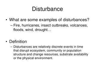
CIRCULATORY DISTURBANCE. Classification:. 1- Disturbance in the volume of circulating blood. 2- Obstruction in the cardiovascular system. 1- Disturbance in Blood volume:. 1- Hyperaemia : increase in the blood flow to an organ as result of dilation of its arterioles and capillaries.

E N D
Classification: • 1- Disturbance in the volume of circulating blood. • 2- Obstruction in the cardiovascular system.
1- Disturbance in Blood volume: 1- Hyperaemia: increase in the blood flow to an organ as result of dilation of its arterioles and capillaries. 2- Congestion: increase venous blood in an organ as result of obstruction to venous outflow, the veins, venules and capillaries becom passively dilated.
1- Disturbance in Blood volume: 3- Haemorrhage: Escape of blood outside the blood vessels or cardiac chambers.
1: Chronic venous congestion in lung: -the alveolar capilleries are dilated and congested ,the alveolar walls are thickened -the alveolar spaces contain intact and haemolysed red cells , brown haemosiderin granules and heart failure cells.
2- Chronic venous congestion in liver: -the central veins are dilated and congested. The sinusoids are dilated and congested, they contain excess red cells haemosiderin and necrotic liver cells.
3- Cerebral Heamorrhage: Cerebral hemorrhage is usually caused by rupture of a small vessel in the parenchyma. The haemorrhage may be the result of an injury, an abnormality of the blood vessels, or high blood pressure.
4- Cerebral Haematoma: haematoma (black), or blood clot,) cerebral hemisphere. This sort of blood clot, which is often fatal, is caused by blood vessels in the brain haemorrhaging..
2- Obstruction of blood flow can be due to: 1-Thrombosis: formation of a compact mass composed of elements of circulating blood inside a vessel or heart cavity, this compact mass is called Thrombus. 2- Clot: a mass of blood elements formed in stagnant blood. 3- Embolism: the process of impaction of embolus in narrow vessels. Embolus is insoluble substance circulating in blood stream.
2- Obstruction of blood flow can be due to: 4-Ischemia: decrease blood supply to apart of tissue due to occlusion of its artery. 5-Infarction:formation of an infarct, that is, an area of tissue death (necrosis) due tosudden ischemia.
5- Thrombosis in the out flow of the heart: showing a blood clot (thrombus, dark red), blood clot can block or reduce the blood supply, will lead to tissue death (infarction)
6- Recent thrombus: • The lumen is occupied by a thrombus appearing as a red mass. • The substance of the thrombus in traversed by lines of Zahn ( fused platelets forming homogenous structure less pinkish violet lines) • .the periphery of the lines show bluish dots represent WBC and nuclear fragments
7- Pulmonary embolism and infarction: - lung showing an area of tissue death (infarction) caused by an embolism. The dead tissue is at lower centre. A pulmonary embolism is a blockage in one of the branches of the pulmonary artery, which carries blood to the lungs to pick up oxygen.
8- Pulmonary infarction: -the alveolar walls in the infarct area are thinned and their cellular structures are lost and fibrous septa only remains. the alveolar spaces contain intact and haemolysed red cells and heart failure cells. - Rest of lung shows CVC.
9- Infarction in kidney: -the infarct is pale red, normal area is darker. - In the infarct zone the normal structures are lost, but the glomerulioutlines, tubules and B.V are homogenous pink shadows
10- Atherosclerosis with lipid foam cells: Atherosclerosis(artery wall thickens, hardening caused by the formation of plaqueswithin the arteries) Lesion initial in intema, have soft yellow accumulaton of lipid ,machrophages, foam cells (macrophages with ingested oxidized LDL),lymphocytes
11- Atheroma in the heart: A theroma:A fatty deposit in the intima(inner lining) of an artery. Appear white to whitish yellow color.
12- Atheroma, Aneurysm in Aorta : Atheroma in aorta lead to Local (dilatation) swelling of artery cause aneurysm is thin The wall of artery and lumin is filled by thrombus
13- Aneurysm in abdominal artery (Aorta): Atheroma in aorta lead to Local (dilatation) swelling of artery cause aneurysm is thin The wall of artery and lumin is filled by thrombus .
14- Renal Atherosclerosis: This is hyperplastic arteriolosclerosis, which most often appears in the kidney in patients with malignant hypertension. The arteriolar wall is markedly thickened and the lumen is narrowed