Download
1 / 47
510 likes | 806 Views
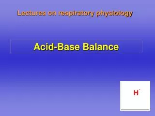
ACID-BASE BALANCE. By: Husnil Kadri Biochemistry Departement Medical Faculty Of Andalas University Padang. Hendersen-Hasselbalch (1909). CARA TRADISIONAL :. HCO 3. HCO 3. [HCO 3 - ]. BASA. GINJAL. Normal. pH = 6.1 + log. Kompensasi. CO 2. pCO 2. PARU. ASAM. CO 2.

E N D
ACID-BASE BALANCE By: HusnilKadri Biochemistry Departement Medical Faculty Of Andalas University Padang
Hendersen-Hasselbalch(1909) CARA TRADISIONAL :
HCO3 HCO3 [HCO3-] BASA GINJAL Normal pH = 6.1 + log Kompensasi CO2 pCO2 PARU ASAM CO2 Normal
Carbonic acid/bicarbonate buffer system pKa = 6.1 • The pKa of carbonic acid is 6.1 • Carbonic acid is the major buffer in ECF • The pH of blood can be determined using the Henderson-Hasselbalch equation H2CO3 H+ + HCO3- ECF: Carbonic acid Bicarbonate ion
Henderson-Hasselbalch equation • pH = pKa + log [HCO3-]/[H2CO3] • pH = pKa + log [HCO3-]/0.03 x PCO2 • 7.4 = 6.1 + log 20 / 1 • 7.4 = 6.1 + 1.3 • Plasma pH equals 7.4 when buffer ratio is 20/1 • The solubility constant of CO2 is 0.03
Normal Compensatory Response • Any primary disturbance in acid-base homeostasis invokes a normal compensatory response. • A primary metabolic disorder leads to respiratory compensation, and a primary respiratory disorder leads to an acute metabolic response due to the buffering capacity of body fluids. • A more chronic compensation (1-2 days) due to alterations in renal function.
Mixed Acid - Base Disorder • Most acid-base disorders result from a single primary disturbance with the normal physiologic compensatory response and are called simple acid-base disorders. • In certain cases, however, particularly in seriously ill patients, two or more different primary disorders may occur simultaneously, resulting in a mixed acid-base disorder. • The net effect of mixed disorders may be additive (eg, metabolic acidosis and respiratory acidosis) and result in extreme alteration of pH; • or they may be opposite (eg, metabolic acidosis and respiratory alkalosis) and nullify each other’s effects on the pH.
Cara Stewart ; DUA VARIABEL pH atau [H+] DALAM PLASMA DITENTUKAN OLEH VARIABEL INDEPENDEN VARIABEL DEPENDEN • Stewart PA. Can J Physiol Pharmacol 61:1444-1461, 1983.
INDEPENDENT VARIABLES DEPENDENT VARIABLES Strong Ions Difference pH pCO2 Protein Concentration
VARIABEL INDEPENDEN CO2 STRONG ION DIFFERENCE WEAK ACID pCO2 SID Atot
DEPENDENT VARIABLES H+ HCO3- OH- AH CO3- A-
CO2 • Rx dominandari CO2adalahrxabsorpsi OH-hasildisosiasi air denganmelepas H+. • Semakintinggi pCO2semakinbanyak H+ yang terbentuk. • Iniygmenjadidasardariterminologi “respiratory acidosis,” yaitupelepasan ion hidrogenakibat pCO2 CO2Didalam plasma beradadalam 4 bentuk • sCO2 (terlarut) • H2CO3asamkarbonat • HCO3- ion bikarbonat • CO32- ion karbonat
STRONG ION DIFFERENCE Definisi: Strong ion difference adalah ketidakseimbangan muatan dari ion-ion kuat. Lebih rinci lagi, SID adalah jumlah konsentrasi basa kation kuat dikurangi jumlah dari konsentrasi asam anion kuat. Untuk definisi ini semua konsentrasi ion-ion diekspresikan dalam ekuivalensi (mEq/L). Semua ion kuat akan terdisosiasi sempurna jika berada didalam larutan, misalnya ion natrium (Na+), atau klorida (Cl-). Karena selalu berdisosiasi ini maka ion-ion kuat tersebut tidak berpartisipasi dalam reaksi-reaksi kimia. Perannya dalam kimia asam basa hanya pada hubungan elektronetraliti.
STRONG ION DIFFERENCE Gamblegram Mg++ Ca++ K+ 4 SID Na+ 140 Cl- 102 [Na+] + [K+] + [kation divalen] - [Cl-] - [asam organik kuat-] [Na+] + [K+] - [Cl-] = [SID] 140 mEq/L + 4 mEq/L - 102 mEq/L = 34 mEq/L KATION ANION
SKETSA HUBUNGAN ANTARA SID,H+ DAN OH- [H+] [OH-] Konsentrasi [H+] Asidosis Alkalosis SID (–) (+) Dalam cairan biologis (plasma) dgn suhu 370C, SID hampir selalu positif, biasanya berkisar 30-40 mEq/Liter
WEAK ACID [Protein-] + [H+] [Protein H] Kombinasi protein dan posfat disebut asam lemah total (total weak acid) [Atot]. Reaksi disosiasinya adalah: disosiasi [Atot] (KA) = [A-].[H+]
Gamblegram Mg++ HCO3- 24 Ca++ K+ 4 SID Na+ 140 Weak acid (Alb-,P-) Cl- 102 KATION ANION
APLIKASI H3O+ = H+ = 40 mEq/L HCO3- HCO3- HCO3- HCO3 = 24 Na 140 K SID Mg SID n Ca SID Alb P Alb Cl 115 Laktat/keto=UA P Alb Cl 102 Cl 102 P CL 95 Asidosis hiperkloremi Keto/laktat asidosis Alkalosis hipokloremi KATION ANION
KLASIFIKASI GANGGUAN KESEIMBANGAN ASAM BASA BERDASARKAN PRINSIP STEWART Fencl V, Jabor A, Kazda A, Figge J. Diagnosis of metabolic acid-base disturbances in critically ill patients. Am J Respir Crit Care Med 2000 Dec;162(6):2246-51
ASIDOSIS ALKALOSIS I. Respiratori PCO2 PCO2 II. Nonrespiratori (metabolik) 1. Gangguan pd SID a. Kelebihan / kekurangan air [Na+], SID [Na+], SID b. Ketidakseimbangan anion kuat: [Cl-], SID [Cl-], SID i. Kelebihan / kekurangan Cl- ii. Ada anion tak terukur [UA-], SID 2. Gangguan pd asam lemah i. Kadar albumin [Alb] [Alb] ii. Kadar posphate [Pi] [Pi] KLASIFIKASI Fencl V, Jabor A, Kazda A, Figge J. Diagnosis of metabolic acid-base disturbances in critically ill patients. Am J Respir Crit Care Med 2000 Dec;162(6):2246-51
RESPIRASI M E T A B O L I K Abnormal pCO2 Abnormal SID Abnormal Weak acid Alb PO4- Anion kuat AIR Cl- UA- Turun Alkalosis Turun kekurangan Hipo Asidosis Meningkat kelebihan Hiper Positif meningkat Fencl V, Am J Respir Crit Care Med 2000 Dec;162(6):2246-51
KEKURANGAN AIR - WATER DEFICIT Diuretic Diabetes Insipidus Evaporasi Plasma Plasma Na+ = 140 mEq/L Cl- = 102 mEq/L SID = 38 mEq/L 140/1/2 = 280 mEq/L 102/1/2= 204 mEq/L SID = 76 mEq/L 1 liter ½ liter SID : 38 76= alkalosis ALKALOSIS KONTRAKSI
KELEBIHAN AIR - WATER EXCESS Plasma 140/2 = 70 mEq/L 102/2 = 51 mEq/L SID = 19 mEq/L Na+ = 140 mEq/L Cl- = 102 mEq/L SID = 38 mEq/L 1 Liter H2O 1 liter 2 liter SID : 38 19= Acidosis ASIDOSIS DILUSI
GANGGUAN PD SID:Pengurangan Cl- Plasma Na+ = 140 mEq/L Cl- = 95 mEq/L SID = 45 mEq/L 2 liter SID ALKALOSIS ALKALOSIS HIPOKLOREMIK
GANGGUAN PD SID:Penambahan/akumulasi Cl- Plasma Na+ = 140 mEq/L Cl- = 120 mEq/L SID = 20 mEq/L 2 liter SID ASIDOSIS ASIDOSIS HIPERKLOREMIK
PLASMA + NaCl 0.9% Plasma NaCl 0.9% Na+ = 140 mEq/L Cl- = 102 mEq/L SID = 38 mEq/L Na+ = 154 mEq/L Cl- = 154 mEq/L SID = 0 mEq/L 1 liter 1 liter SID : 38
ASIDOSIS HIPERKLOREMIK AKIBAT PEMBERIAN LARUTAN Na Cl 0.9% Plasma = Na+ = (140+154)/2 mEq/L= 147 mEq/L Cl- = (102+ 154)/2 mEq/L= 128 mEq/L 2 liter SID = 19 mEq/L SID : 19 Asidosis
PLASMA + Larutan RINGER LACTATE Plasma Ringer laktat Laktat cepat dimetabolisme Na+ = 140 mEq/L Cl- = 102 mEq/L SID= 38 mEq/L Cation+ = 137 mEq/L Cl- = 109 mEq/L Laktat- = 28 mEq/L SID = 0 mEq/L 1 liter 1 liter SID : 38
Normal pH setelah pemberian RINGER LACTATE Plasma = Na+ = (140+137)/2 mEq/L= 139 mEq/L Cl- = (102+ 109)/2 mEq/L = 105 mEq/L Laktat- (termetabolisme) = 0 mEq/L 2 liter SID = 34 mEq/L SID : 34 lebih alkalosis dibanding jika diberikan NaCl 0.9%
MEKANISME PEMBERIAN NA-BIKARBONAT PADA ASIDOSIS Plasma; asidosis hiperkloremik Plasma + NaHCO3 Na+ = 140 mEq/L Cl- = 130 mEq/L SID =10 mEq/L Na+ = 165 mEq/L Cl- = 130 mEq/L SID = 35 mEq/L 25 mEq NaHCO3 HCO3 cepat dimetabolisme 1 liter 1.025 liter SID : 10 35 : Alkalosis, pH kembali normal namun mekanismenya bukan karena pemberian HCO3- melainkan karena pemberian Na+ tanpa anion kuat yg tidak dimetabolisme seperti Cl- sehingga SID alkalosis
UA = Unmeasured Anion: Laktat, acetoacetate, salisilat, metanol dll. Na+ HCO3- HCO3- SID Na+ K K SID Keto- A- A- Cl- Cl- Lactic/Keto asidosis Normal Ketosis
GANGGUAN PD ASAM LEMAH: Hipo/Hiperalbumin- atau P- Na HCO3 Na HCO3 HCO3 Na K K K SID SID SID Alb-/P- Alb-/P- Alb/P Cl Cl Cl Asidosis hiperprotein/ hiperposfatemi Alkalosis hipoalbumin/hipoposfatemi Normal Acidosis Alkalosis
Anion Gap • Described by Gamble in 1939 • Electroneutrality • Na+, Cl-, and HCO3 are measured ions Na + UC = Cl + HCO3 + UA UC= Sum of unmeasured cations UA = Sum of unmeasured anions
Unmeasured Cations: total 11 mEq/L Potassium 4 Calcium 5 Magnesium 2 Unmeasured Anions: total 23 mEq/L Sulfates 1 Phosphates 2 Albumin 16 Lactic acid 1 Org. acids 3 Anion Gap
Anion Gap Na + UC = Cl + HCO3 + UA 140 + 11 = 104 + 24 + 23 151 = 151 UA – UC = Na - (Cl + HCO3); Anion Gap = Na - (Cl + HCO3)
If the anion gap is elevated • Then compare the changes from normal between the anion gap and [HCO3 -]. • If the change in the anion gap is greater than the change in the [HCO3 -] from normal, then a metabolic alkalosis is present in addition to a gap metabolic acidosis. • If the change in the anion gap is less than the change in the [HCO3 -] from normal, then a non gap metabolic acidosis is present in addition to a gap metabolic acidosis.
Anion Gap Acidosis: • Anion gap >12 mEq/L; caused by a decrease in [HCO3 -] • balanced by an increase in an unmeasured acid ion from either endogenous production or exogenous ingestion (normochloremic acidosis).
Non anion Gap Acidosis: • Anion gap = 8-12 mmol/L; caused by a decrease in [HCO3 -] balanced by an increase in chloride (hyperchloremic acidosis). Renal tubular acidosis is a type of non gap acidosis
Increased Anion GapNormal = 8-15May differ institutionally • Accumulation of organic acids (ketones, lactate) • Toxic Ingestions • methanol, ethylene glycol, salicylates • Reduced inorganic acid excretion • phosphates, sulfates • Decrease in unmeasured cations (unusual)
Methanol Uremia/Renal Failure INH, Iron--lactate Paraldehyde Lactic Acidosis Has many etiologies Cyanide, CO, Toluene, HS Poor perfusion Ethylene glycol Salicylates Methyl salicylate (Oil of wintergreen) Mg salicylate Increased AG Metabolic Acidosis: Levraut J et al. Int Care Med 23:417, 1997
Increased Anion GapNormal = 8-15May differ institutionally“ion specific electrodes” • Accumulation of organic acids (ketones, lactate) • Toxic Ingestions • methanol, ethylene glycol, salicylates • Reduced inorganic acid excretion • phosphates, sulfates • Decrease in unmeasured cations (unusual)
Decreased or Negative Anion GapClin J Am Soc Nephrol 2: 162-174, 2007 • Low protein most important • Albumin has many unmeasured negative charges • “Normal” anion gap (12) in cachectic person • Indicates anion gap metabolic acidosis • Other etiologies of low AG: • Low K, Mg, Ca, increased globulins (Mult. Myeloma), I intoxication • Negative AG • more unmeasured cations than unmeasured anions • Bromide, Iodide, Multiple Myeloma
Change in Anion Gap vs HCO3 • In simple AG Metabolic Acidosis • decrease in plasma bicarbonate = increase in AG Anion Gap = 1 HCO3 • Helpful in identifying mixed disorders
Metabolic Acidosis: Occurs rapidly Hyperventilation “Kussmaul Respirations” Deep > rapid (high tidal volume) Is not Respiratory Alkalosis Metabolic Alkalosis: Calculation not as accurate Hypoventilation Not Respiratory Acidosis Restricted by hypoxemia PCO2 seldom > 50-55 Respiratory Compensationfor
Reference • Achmadi, A., George, YWH., Mustafa, I. Pendekatan “Stewart” DalamFisiologiKeseimbanganAsamBasa. 2007 • Beaudoin, D. Electrolytes and ion sensitive electrodes. PPT. 2003. • Ivkovic, A ., Dave, R. Renal review. PPT • Kersten. Fluid and electrolytes. PPT. • Marieb, EN. Fluid, electrolyte, and acid-base balance. PPT. Pearson Education, Inc. 2004 • Rashid, FA. Respiratory mechanism in acid-base homeostasis. PPT. 2005. • Silverthorn, DU. Integrative Physiology II: Fluid and Electrolyte Balance. Chapter 20, part B. Pearson Education, Inc. 2004 • Smith, SW. Acid-Base Disorders. www.acid-base.com