Download
1 / 38
421 likes | 780 Views
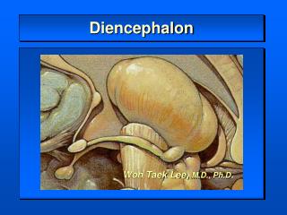
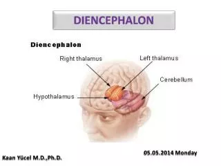
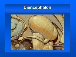
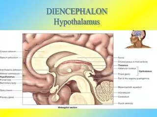
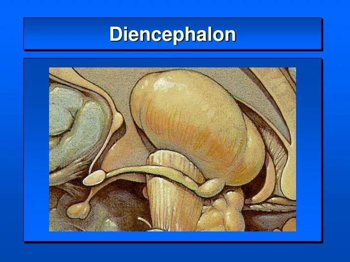
Diencephalon. Diencephalon. Thalamus dorsal thalamus Hypothalamus pituitary gland Epithalamus habenular nucleus and commissure pineal gland Subthalamus ventral thalamus subthalamic nucleus (STN) field of Forel . Diencephalon. dorsal surface. Diencephalon.

E N D
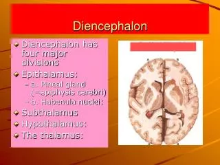
Diencephalon • Thalamus dorsal thalamus • Hypothalamus • pituitary gland • Epithalamus • habenular nucleus and commissure • pineal gland • Subthalamus ventral thalamus • subthalamic nucleus (STN) • field of Forel
Diencephalon dorsal surface
Diencephalon ventral surface
Diencephalon Medial Surface
Classification of Thalamic Nuclei I. Lateral Nuclear Group II. Medial Nuclear Group III. Anterior Nuclear Group IV. Posterior Nuclear Group V. Metathalamic Nuclear Group VI. Intralaminar Nuclear Group VII. Thalamic Reticular Nucleus
LATERAL NUCLEAR GROUP • Ventral Nuclear Group • Ventral Posterior Nucleus (VP) • ventral posterolateral nucleus • ventral posteromedial nucleus (VPM) • ventral posteroinferior nucleus (VPI)
LATERAL NUCLEAR GROUP Ventral Lateral Nucleus (VL) Ventral Anterior Nucleus (VA, L.po.)
LATERAL NUCLEAR GROUP Prefrontal SMA MI, PM SI Ventral Nuclear Group TTT SNr GPi Cbll ML, STT
LATERAL NUCLEAR GROUP Dorsal or Lateral Nuclear Group Lateral Dorsal Nucleus (LD, D.sf.) Lateral Posterior Nucleus (LP, D.im. & Z.im.) Pulvinar (P, Pu.)
LATERAL NUCLEAR GROUP cingulate gyrus, precuneus Somesthetic Association Area Visual Association area HF SC, Pretectal
MEDIAL NUCLEAR GROUP Dorsomedial Nucleus (MD) - pars magnocellularis - pars parvocellularis Midline Nuclear Group
ANTERIOR NUCLEAR GROUP • anteroventral nucleus (AV, A.pr.) • anterodorsal nucleus (AD, A.d.) • anteromedial nucleus (AM, A.m.) • POSTERIOR NUCLEAR GROUP • suprageniculate nucleus (SG, Li.) • nucleus limitans (Lim, Li.) • posterior nucleus (PO, Li.por.)
MEDIAL & ANTERIOR NUCLEAR GROUP Prefrontal Cortex Frontal Eye Field MB, HF Medial Frontal Gyrus cingulate gyrus Basal forebrain SNr, SC, RF
METATHALAMIC NUCLEAR GROUP • Medial Geniculate Nucleus (MG) • ventral nucleus • dorsal nucleus • medial nucleus • Lateral Geniculate Nucleus (LG) • dorsal nucleus • ventral nucleus
INTRALAMINAR NUCLEAR GROUP • Rostral Intralaminar Nuclei • central lateral nucleus, • central medial nucleus, • paracentral nucleus, • Caudal Intralaminar Nuclei • (CM-PF nuclear complex) • centre median nucleus, • parafascicular nucleus,
THALAMIC RETICULAR NUCLEUS Cerebral Cortex Thalamocortical Neuron Thalamic Reticular Nucleus Subcortical Structure
Summary of Thalamic Connectivity I. Sensory Input general sensation special sensation taste, equilibrium, hearing, vision II. Motor Input cerebellum, basal ganglia III. Reticular Formation IV. Limbic System mammillary nucleus hippocampal formation
Sensory Input (1) General Sensation 1. Medial lemniscus VPLc ----------- S I, S II 2. Spinothalamic tract VPLc ----------- S I, S II POm ----------- retroinsular cortex Sm ------------- frontal lobe(?) CL ------------- diffuse cortical areas 3. Trigeminothalamic tract VPM ------------- S I, S II
Sensory Input (2) Taste sensation VPM pc ------- gustatory area (Brodmann area 43) (3) Sense of equilibrium VPLo ---------- S I (4) Auditory sensation MGv ----------- A I (Brodmann area 41, 42) MGd ----------- A II (5) Vision LGd ---------- V I (Brodmann area 17) Pi, Pl ---------- V I, V II (Brodmann area 17, 18, 19)
Motor Input (1) Cerebellum VLc, VPLo, - M I (Brodmann area 4), Premotor area (2) Basal ganglia 1. GPi (internal segment of globus pallidus) -------- SMA (supplementary motor area) ------- frontal cortex 2. SNr (pars reticularis of substantia nigra) - fronal cortex --------- medial frontal cortex ---------- frontal eye field (Brodmann area 8)
Reticular Formation & Limbic System Reticular formation rostral intralaminar nuclei (CL, Cem, PC), Rh ----- diffuse cortical areas thalamic reticular nucleus (R), LGv ----- adjacent thalamic nucleus Limbic System Mammillary Body AV, AD, AM ----- cingulate gyrus Hippocampal Formation AV, AD, AM ----- cingulate gyrus LD ------------------ cingulate gyrus, precuneus Re ------------------- medial cortex, entorhinal cortex
Clinical Syndromes of the Thalamus Posterolateral thalamic syndromes sensory disorders Thalamic (Dejerine-Roussy) syndrome ----- VP nucleus - pain Medial thalamic syndromes disorders of consciousness thalamic neglect, thalamic amnesia, akinetic mutism Anterolateral thalamic syndromes motor disorders paresis, ataxia, motor incoordination, dysphagia
Thalamic (Dejerine-Roussy) Syndrome Joseph Jules Dejerine (1849-1917) Gustave Roussy (1874-1948)
Visual (Optic) Pathway Modality: Vision Receptor:Photoreceptor Cell of Retina Cranial Nerve: II (Optic nerve) 1st Neuron:Bipolar Cell 2nd Neuron:Ganglion Cell optic nerve optic chiasm optic tract 3rd Neuron:Lateral Geniculate Nucleus optic radiation Termination:Visual Areas (V I, V II) Brodmann area 17 (V I), 18, 19 (V II)
RETINA - Pars Nervosa 1. Pigment Epithelium - retinal detachment 2. Neuronal Layer (1) Photosensitive Cell Layer Rod Cell, Cone Cell (2) Bipolar Cell Layer Bipolar Cell Horizontal Cell Amacrine Cell (3) Ganglion Cell Layer Ganglion Cell Optic Nerve (II)
Visual Pathway 1. Optic nerve 2. Optic chiasm 3. Optic tract 4. Lateral geniculate body 5. Optic radiation 6. Visual cortex
Visual PathwayLateral Geniculate Nucleus (LGd) Dorsal Nucleus (LGd) Magnocellular Part 1, 2 Parvocellular Part 3, 4, 5, 6 contralateral afferents 1, 4, 6 ipsilateral afferents 2, 3, 5 Ventral Nucleus (LGv) part of thalamic reticular nucleus dorsolateral ventromedial
Optic Radiation (Geniculocalcarine Tract) Meyer’s loop
Clinical Features of Visual Pathway Lesion 1. optic nerve 2. optic chiasm 3. optic tract 4. 5. optic radiation A. unilateral blindness B. bitemporal hemianopsia C. left homonymous hemianopsia D. left inferior homony- mous quadranopsia E. left superior homony- mous quadranopsia
Signs of Visual Pathway Lesion Optic nerve - ipsilateral blindness Optic chiasm - bitemporal hemianopsia Optic tract - contralateral homonymous hemianopsia Optic radiation - contralateral homonymous quadranopsia - intact light reflex Visual Cortex - contralateral homonymous quadranopsia - macular sparing
Visual Field Defect left inferior optic radiation lesion right superior quadranopsia
Epithalamus Limbic System Habenular Nucleus Medial Habenular Nucleus Lateral Habenular Nucleus Habenular Commissure Pineal Gland
Subthalamus Basal Ganglia Subthalamic nucleus zona incerta Field of Forel H ansa lenticularis H1 thalamic fasciculus H2 lenticular fasciculus