Download
1 / 41
410 likes | 539 Views
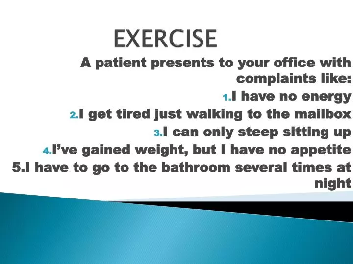
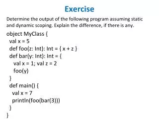
EXERCISE. A patient presents to your office with complaints like: I have no energy I get tired just walking to the mailbox I can only steep sitting up I’ve gained weight, but I have no appetite 5.I have to go to the bathroom several times at night. CONGESTIVE HEART FAILURE. BY

E N D
EXERCISE A patient presents to your office with complaints like: I have no energy I get tired just walking to the mailbox I can only steep sitting up I’ve gained weight, but I have no appetite 5.I have to go to the bathroom several times at night
CONGESTIVE HEART FAILURE BY LEBURA EWONUBARI GRACE ROLL NO: 666
OUTLINE • DEFINITION • ETIOLOGY/CAUSES • EPIDEMIOLOGY • PATHOGENESIS • PATHOPHYSIOLOGY • SIGNS AND SYMPTOMS • RISK FACTORS • CLASSIFICATION:STAGES/TYPES • DIAGNOSIS • PROGNOSIS • COMPLICATIONS • PREVENTION • TREATMENT • SUMMARY • REFERENCES
DEFINITION • The term Congestive Heart failure • or cardiac failure means simply • failure of the heart to • pump enough blood to satisfy • the needs of the body
ETIOLOGY/CAUSES • Myocardial infarction • Viral myocarditis • Coronary artery disease • Long term hypertension • Infiltrations of the muscle such as amyloidosis
EPIDEMIOLOGY • leading cause of hospitalization • In people older than 65 • 6.6million people in the US • 23 milion people worldwide. • Men have a higher incidence • women survive longer
PATHOGENESIS • (1) reduced cardiac output and • (2) volume overload due to damming • of blood in the veins, • resulting in increased venous pressure.
PATHOPHYSIOLOGY • Reduced force of contraction, due • to overloading of the ventricle • A reduced stroke volume, due to • failure of systole, diastole or both • Increased heart rate, • stimulated by increased sympathetic activity • in order to maintain cardiac output.
SIGNS AND SYMPTOMS • Shortness of breath • Fatigue and weakness • Swelling (edema) in your legs • Increased need to urinate at night • Swelling of abdomen (ascites) • Sudden weight gain • Elevated blood pressure
RISK FACTORS Age • Hypertension • Diabetes • Obesity • Coronary artery disease • High cholesterol and triglycerides • Prior heart attack
STAGES • The American College of Cardiology/ • American Heart Association stages patients according to the progression of • their heart failure: • Stage A: High risk for developing heart failure • Stage B: Asymptomatic heart failure • Stage C: Symptomatic heart failure • Stage D: Refractory end-stage heart failure
The New York Heart Association • classifies patients based on their • physical limitations. Classifications are as follows: • Class I: No limitations of physical activity • Class II: Slight limitation • Class III: Marked limitation • Class IV: Severe limitation
TYPESLEFT SIDED FAILURE • Due to Decreased ventricular contraction (systolic dysfunction) • Causes of systolic dysfunction: • Ischemia due to coronary artery atherosclerosis • Post-myocardial infarction, myocardial fibrosis, myocarditis, dilated cardiomyopathy • Noncompliantventricle (diastolicdysfunction) • Restricted filling of the left ventricle • in diastole due to essential hypertension
Clinical features: • Pulmonary congestion leads to pulmonary edema. • i. Results in dyspnea, paroxysmal nocturnal dyspnea, orthopnea, and crackles • Decreased flow to kidneys leads • to activation of renin-angiotensin system Fluid retention exacerbates CHF
RIGHT SIDED FAILURE • Most commonly due to • left-sided heart failure • left to right shunt and • chronic lung disease (corpulmonale). • Due to: Decreased contraction • b. Noncompliant right ventricle • c. Increased afterload
Clinical features are due to congestion. • 1. Jugular venous distension • 2. Painful hepatosplenomegaly with characteristic 'nutmeg' liver , may lead to cardiac cirrhosis • 3. Dependent pitting edema (due to increased hydrostatic pressure)
SYSTOLIC FAILURE • stroke volume is reduced because • ventricular contraction is weak. • the ejection fraction falls from • 65% to as low as 20%. • The initial response to failure • is activation of the genes that cause cardiac myocytes to hypertrophy, • and thicken of the ventricular wall (cardiac remodeling).
DIASTOLIC FAILURE • the ejection fraction is initially • maintained but the elasticity of the myocardium is reduced so filling during diastole is reduced. • This leads to inadequate stroke volume • and the same cardiac remodeling • and Na' and water retention • that occur in systolic failure.
HIGH OUTPUT FAILURE • cardiac output is increased compared • with values for the normal resting state, • due to:. Increase in stroke volume . (Eg hyperthyroidism) • Decrease in blood viscosity • Decrease total peripheral resistance (TPR) • Increase venous return to the heart. (Egsevere anemia)
LOW OUTPUT FAILURE • After acute heart attacks and • often after prolonged periods of • slow progressive cardiac deterioration, the heart becomes incapable of pumping • even the minimal amount of blood flow required to keep the body alive. • Consequently, all the body tissues • begin to suffer and even to deteriorate, • often leading to death within a few hours to a few days.
PHYSICAL EXAMINATION • In mild or moderately severe HF, • the patient appears to be • in no distress at rest except • for feeling uncomfortable • In more severe HF, • the patient must sit upright, • may have labored breathing
Examination of the jugular veins • provides an estimation of right atrial pressure. • Pulmonary crackles (rales or crepitations) result from the transudation of • fluid from the intravascular space • into the alveoli. • In patients with pulmonary edema, • ralesmay be heard widely over both lung fields
In some patients, a third • heart sound (S3) is audible • and palpable at the apex. • Patients with enlarged or hypertrophied • right ventricles may have a sustained • and prolonged left parasternal impulse extending throughout systole.
Hepatomegaly is an important sign • Ascites, a late sign, • occurs as a consequence of increased pressure in the hepatic veins and • the veins draining the peritoneum. • Jaundice, also a late finding in HF, • results from impairment of hepatic • function secondary to hepatic congestion
DIAGNOSIS • Routine Laboratory Testing • Patients with new-onset HF • and those with chronic HF • and acute decompensation should have • a complete blood count, a panel of electrolytes, blood urea nitrogen, serum creatinine, hepatic enzymes, and a urinalysis.
Electrocardiogram (ECG) • The major importance of the ECG • is to assess cardiac rhythm and • determine the presence of LV hypertrophy • or a prior MI (presence or absence of Q waves
Chest X-Ray • A chest x-ray provides useful information about cardiac size and shape, • as well as the state • of the pulmonary vasculature, and • may identify noncardiac causes of • the patient's symptoms.
Assessment of LV Function • Noninvasive cardiac imaging is essential • for the diagnosis, evaluation, and management of HF.
Biomarkers • Both B-type natriuretic peptide (BNP) • and N-terminal pro-BNP, which are • released from the failing heart, • are relatively sensitive markers for the presence of HF with depressed EF
they also are elevated in HF • patients with a preserved EF, • albeit to a lesser degree. • Other biomarkers, such as troponin T • and I, C-reactive protein, TNF receptors, • and uric acid, may be elevated in HF.
Exercise Testing • Treadmill or bicycle exercise testing • is not routinely advocated for • patients with HF, but either • is useful for assessing the need • for cardiac transplantation in patients • with advanced HF .
PROGNOSIS • In general, the mortality following hospitalization for patients with heart • failure is 10.4% at 30 days, • 22% at 1 year, and 42.3% at 5 years, • despite marked improvement in medical • and device therapy. • Each rehospitalization increases mortality by about 20-22%.
COMPLICATIONS • Kidney damage or failure • Heart valve problems • Liver damage • Stroke:Because blood flow through • the heart is slower in heart failure • than in a normal heart, blood clots can develop.
PREVENTION • control or eliminate many of • the risk factors for heart disease —: • high blood pressure and coronary artery disease, for example. • Not smoking • Controlling certain conditions, such as • high blood pressure, high cholesterol and diabetes • Staying physically active
TREATMENT • ACE INHIBITORS • ANGIOTENSIN RECEPTOR BLOCKERS(ARBs) • BETA BLOCKERS • DIURETICS • For valve problem: valve repair or replacement is done. • Coronary artery bypass graft(CABG)
Angioplasty (open up closed artery) • Severe cases may require surgical • implants such as pacemakers, ventricular assist device (VAD).
SUMMARY • CHF occurs when the heart • is unable to pump blood at • a rate that meets the metabolic • requirements of the peripheral tissue; • inadequate cardiac output is usually accompanied by increased congestion of • the relevant venous circulation.
Left-sided heart failure is most • commonly due to IHD, • systemic hypertension, mitral or aortic valve disease, and primary diseases of the myocardium; symptoms are primarily related to pulmonary congestion and edema.
Right-sided heart failure is • most commonly due to left-sided • heart failure or to primary pulmonary diseases; • it is associated with peripheral • edema and visceral congestion.
REFERENCES • Arthur S, Philip A.(2002). The Heart: Congestive Heart Failure. Board Review Series pathology, 2nd Edition. 144 – 145. • Edward G.(2010): Heart Disorders: Rapid Review Pathology, 3rd Edition. 158 – 164. • Guyton and Hall (2006). Cardiac Failure. Textbook of Medical Physiology, Eleventh Edition.258-268. • Henry S, John B.(2010). Cardiac Pathology: Congestive Heart failure. Kaplan Medical USMLE Step1. 144-145 • Husain A.(2011). Cardiac Pathology: congestive heart failure. Fundamentals of Pathology, Medical Course and step 1 Review, First Edition. 76 • Kumar et al(2007). The Heart:heart failure. Robbins Basic Pathology, 8th Edition. • Longo et al(2012). Disorders of Cardiovascular system: disorders of the heart. Harrison’s Principles of Internal Medicine, 18th Edition. • Stephen J.M and Gary D.H.(2010). Cardiovascular disorders:Heart disease. Pathophysiology of Disease, 6th Edition. • William F. G. (2005). Cardiovascular Homeostasis in Health and Disease: Heart Failure. Review of Medical Physiology, Twenty -Second Edition. 643- 644.

![[Exercise Name] Functional Exercise](https://cdn0.slideserve.com/621913/exercise-name-functional-exercise-dt.jpg)
![[Exercise Name] Functional Exercise](https://cdn1.slideserve.com/1717560/exercise-name-functional-exercise-dt.jpg)


![[Exercise Name] Functional Exercise](https://cdn3.slideserve.com/6680259/exercise-name-functional-exercise-dt.jpg)

![[Exercise Name] Tabletop Exercise](https://cdn4.slideserve.com/9191716/exercise-name-tabletop-exercise-dt.jpg)
![[Exercise Name] Functional Exercise](https://cdn5.slideserve.com/9609621/exercise-name-functional-exercise-dt.jpg)