Download

1 / 12
140 likes | 209 Views
Mycoplasma and Ureaplasma. Dr. Hala Al Daghistani. MYCOPLASMA. Smallest free-living micro organisms, lack cell wall. Many can pass through a bacterial filter. Called “pleuropneumonia like organisms”(PPLO). 1956- PPLO replaced by Mycoplasma. Myco : fungus like branching filaments

E N D
Mycoplasma and Ureaplasma Dr. Hala Al Daghistani
MYCOPLASMA • Smallestfree-living micro organisms, lack cell wall. • Many can pass through a bacterial filter. • Called “pleuropneumonia like organisms”(PPLO). • 1956- PPLO replaced by Mycoplasma. • Myco : fungus like branching filaments • Plasma : plasticity highly pleomorphic – no fixed shape or size .
Morphology and Physiology • Small genome size (M. pneumoniae is ~800 Kbp) • Require complex media for growth including sterols • Facultative anaerobes • Except M. pneumoniae - strict aerobe • are resistant to penicillins, cephalosporins and vancomycin, etc. • Grow slowly and reproduced by binary fission • Doubling time can be as long as 16 -18hours, extended incubation needed
Mycoplasmas of Humans • Parasitic • Established pathogens: M. pneumonia • Presumed pathogens: M. hominis, U. urealyticum • Non pathogenic: M. orale, M. buccale, M. genitalium, M. fermentans • Saprophytic – present mainly on skin & in mouth.
Pathogenicity Produce surface infections – adhere to the mucosa of respiratory, gastrointestinal & genitourinary tracts with the help of adhesin. Two types of diseases: • Atypical Pneumonia • Genital infections
Adherence • P1 pili (M. pneumoniae) • Toxic metabolic products • Peroxide and superoxide • Inhibition of catalase produced by WBCs • Immunopathogenesis • Stimulate cytokine production • Inflammatory cells migrate to infection and release TNF-a then IL-1 and IL-6
Mycoplasma pneumoniae • Also called Primary Atypical Pneumonia/ Walking pneumonia. • Seen in all ages • Incubation period: 1-3 weeks • Transmission: airbornedroplets of nasopharyngeal secretions, close contacts (families, military recruits). • Gradual onset with fever, malaise, chills, headache & sore throat. • Severe cough with blood tinged sputum • Complications: otitis media, meningitis, encephalitis, hemolytic anemia
Diseases Caused by Mycoplasma Organism Disease M. pneumoniae Atypical pneumonia , Upper respiratory tract disease, tracheobronchitis M. hominis Genital infection, Pyelonephritis, pelvic inflammatory disease, postpartum fever M. genitaliumNongonococcalurethritis U. urealyticumNongonococcalurethritis, (pneumonia and chronic lung disease in premature infants) Both M. hominis, U. urealyticumAlso associated with infertility, abortion, postpartum fever, chorioamnionitis & low birth weight infants
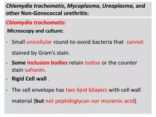
Laboratory Diagnosis • Specimens – throat swabs, respiratory secretions. • Highly pleomorphic • Gram negative, but better stained with Giemsa, Dienes’ stain, • Isolation of Mycoplasma (Culture) • Enriched medium containing 20% horse or human serum (as a source of cholesterol & other lipids) • Incubate aerobically for 7 -12 days with 5–10% CO2at 35-37°C • Typical “fried egg” appearance of colonies - Central opaque granular area of growth extending into the depth of the medium, surrounded by a translucent peripheral zone. 4. Produce beta hemolytic colonies
Fried egg colonies Dr Ekta,Microbiology, GMCA
Identification of Isolates • Growth Inhibition Test– inhibition of growth around discs impregnated with specific antisera. • Immunofluorescence • Molecular diagnosis • Non specific serological tests – cold agglutination tests (Abs agglutinate human group O red cells at low temperature, 4C). 1:32 titer or above is significant.