Download
1 / 1
10 likes | 134 Views
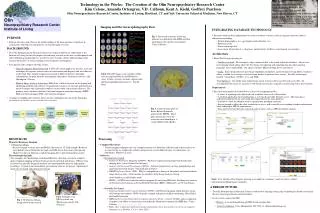
Fig 6. User interface flow diagram showing an example of a summary search to retrieve subject information including image summaries. NRC STAFF Director: Godfrey Pearlson, MD CCNLab Director: Kent Kiehl, PhD MIALab Director: Vince Calhoun, PhD

E N D
Fig 6. User interface flow diagram showing an example of a summary search to retrieve subject information including image summaries. NRC STAFF Director: Godfrey Pearlson, MD CCNLab Director: Kent Kiehl, PhD MIALab Director: Vince Calhoun, PhD IOL Psychiatrist-In-Chief: Harold I. Schwartz, MD Lab Personnel: Mike Stevens PhD, Jinsuh Kim MD, Kristen Mckeirnan PhD, Karen Anderson RN, David Dean, Tim O’Keefe, Karyn Groth, Thania Benios, Amanda Ortengren, Kim Celone, Corey Blumenfeld. Olin Neuropsychiatry Research Center Institute of Living Technology in the Works: The Creation of the Olin Neuropsychiatry Research Center Kim Celone, Amanda Ortengren, V.D. Calhoun, Kent A. Kiehl, Godfrey PearlsonOlin Neuropsychiatry Research Center, Institute of Living, Hartford, CT and Yale University School of Medicine, New Haven, CT Imaging and Electroencephalography Data • INTEGRATING DATABASE TECHNOLOGY • Recently work has been implemented to create a database system in order to organize and store subject information including: • Patient demographics (i.e., age height, medical history, etc.) • Behavioral data • Processed image data • Assessment information (i.e., diagnosis, family history of illness, neurological assessments). • Architecture • Three-Tier Design consisting of: • Database backend: We anticipate a large volume of data to be stored within the database. Also we are investigating which option offers the best means of importing and exporting data into other analysis programs, for example SPSS. Our choices include: Microsoft SQL Server and Oracle. • Logic: Since image retrieval and storage algorithms incorporate a great deal processing ability, we plan to utilize a middle layer of logic to increase time needed to perform these actions. Possible technologies include: Visual Basic (COM+), C++, and XML. • User Interface: One of the main requirements of our system is that it can be easily accessible. To accommodate this need, we envision a web-based user interface typically created using Active Server Pages. • Requirements • The main functionality described above is also to be complemented by: • A series of reporting tools which will aid in further analysis of collected data. • Adesktop application for recording data at sites that do not offer Internet access. This data that is collected would need to be stored and then imported into the main database. • Scalable to allow the addition of new experimental paradigms and data. • Secure enough to allow the other institutions to access and eventually even combine database information while adhering to HIPAA requirements. • Data must be extractable into statistical analysis tools, such as SPSS for further analysis. PURPOSE • Educate ourselves and others in the understanding of the brain and how it functions in conjunction with behavior and genetics via neuroimaging research. • BACKGROUND • The Olin Neuropsychiatry Research Center was made possible by an endowment to the Institute of Living for the sole purpose of furthering research in the area of schizophrenia and other debilitating mental illnesses in order to move towards a better understanding of the diseases themselves as well as finding newer treatments and diagnoses. • Two primary labs comprise the Olin Center: • Clinical Cognitive Neuroscience lab (CCN Lab), which applies the theories, tools and techniques of cognitive neuroscience to better understand diagnose and treat illnesses of the brain. This includes looking at a myriad of different diseases including schizophrenia, bi-polar disorder, psychopathy, drug abuse, Alzheimer's disease, and Huntington’s Chorea. • Medical Image Analysis Laboratory (MIA Lab), which is focused on developing and optimizing methods and software for quantitative analysis of structure and function in medical images with a particular emphasis on the study of psychiatric illnesses. The primary areas of interests include; functional magnetic resonance imaging (fMRI), diffusion tensor imaging (DTI), and structural imaging. • A unique overlapping lab structure allows for the combination of each of the biomarker technologies to produce more scientifically valid data: Fig 3. The neural correlates of driving behavior are identified with fMRI and their modulation with speed is investigated. (Vince Calhoun) Fig 4. This DTI image is an example of fiber tract tracing from Broca's and Wernicke's areas. It shows arcuate, uncinate, superior and inferior longitudinal fasciculi, etc. (Jinsuh Kim) Fig. 5. Grand Average plots of Event-Related Potentials generated by ERPSS. These plots specifically show the activation in relationship to a visual oddball study.(Kiehl) • Processing • Computer Hardware: • Neuro-imaging techniques are very computer intensive in both data collection and in data analysis to account for this we employ the stability and power of several different types of workstations (i.e. Sun/Solaris, Windows, Linux). • Software: • Externally Developed: • Statistical Parametric Mapping (SPM99) - Written to organize and interpret functional neuro-imaging data. (www.fil.ion.ucl.ac.uk/spm/) • Analysis of Functional NeuroImages (AFNI) – Software program for viewing, manipulating, and analyzing functional images. (afni.nimh.nih.gov/afni/) • FMRIB Software Library (FSL) - FSL is a comprehensive library of functional and structural brain image analysis tools, written mainly by members of the Image Analysis Group (www.fmrib.ox.ac.uk/fsl/index.html) • ERPSS - A UNIX based software analysis program for analyzing EEG data. Facilitates the analysis of electrical activity known as Event-Related Potentials (ERPs), (Hillyard et. al, UC San Diego). • Internally Developed: • Visual and Auditory Presentation Package (VAPP): A DOS based program which presents visual (tiff, bitmap) and auditory (WAV;cd quality) stimuli with precise millisecond timing. (Kent Kiehl, kent.kiehl@yale.edu) • fMRI Analysis using Independent Component Analysis (ICA) – A novel, flexible analysis approach to improve our ability to characterize and utilize the information contained in fMRI data, (Vince Calhoun, vince.calhoun@yale.edu) • Many other scripts and utilities for image analysis have been developed in a variety of computer languages (e.g. Matlab, C, C++, Java, IDL) • RESOURCES • Magnetic Resonance Imaging • 3T Seimens Allegra • Increased signal-to-noise ratio and BOLD-effect due to 3 T field strength. Reduced susceptibility-based distortions in single-shot EPI due to ultra-short echo-spacing. Highest b-values in diffusion weighted imaging due to powerful gradient system. • Electroencephalography • Two complete 64 Channel high resolution EEG data collection system for complete spatio-temporal mapping of brain electrical activity in health and disease. EEG systems are housed in specially designed shielded and sound-proofed rooms for high quality recordings. Custom visual and auditory presentation software for precise experimental control of all stimuli and participant behavior • A BRIGHT FUTURE • The Olin Neuropsychiatry Research Center is dedicated to utilizing cutting edge technology to further research in the area of neuropsychiatric mental disorders. • If you wish to contact the NRC: • Website: www.instituteofliving.org/NRC/external_index.htm • Center Coordinator: Corey Blumenfeld: 545-7678 or cblumen@harthosp.org Fig 2. Example of an EEG electrode cap. (Electrocap worn by Tim O’Keefe) Fig 1.3T Seimens Allegra, designed for brain research