Download
1 / 41
430 likes | 628 Views
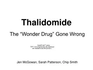
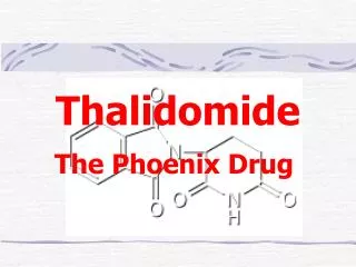
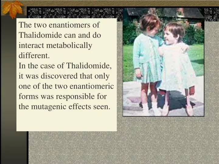
The two enantiomers of Thalidomide can and do interact metabolically different. In the case of Thalidomide, it was discovered that only one of the two enantiomeric forms was responsible for the mutagenic effects seen. Chapter 5 The Structure and Function of Macromolecules.

E N D
The two enantiomers of Thalidomide can and do interact metabolically different. In the case of Thalidomide, it was discovered that only one of the two enantiomeric forms was responsible for the mutagenic effects seen.
Chapter 5The Structure and Function of Macromolecules Polymers: carbohydrates lipids proteins nucleic acids Their structures, sources, uses
Polymers • polys (many) meris (parts) • Built of monomers (single units) • monosaccharides • Amino acids • Nucleotides
Condensation (Dehydration) reaction:builds polymers (ex. on next slide) a water molecule is “made”(-H) (-OH) from the site where to two bond. • Hydrolysis: breaks polymers are disassembled hydro (water) lysis (break) water is broken (-H) (-OH) to fill the “gaps” left when the two parts separate • See fig. 5.2
Condensation= builds longer molecules, H2O results Hydrolysis= breaks H2O bonds, shortens molecules • FIG 5.2
Carbohydrates • mono-, di-, and polysaccharides • CH2O (basic formula) • Carbonyl group (C=O) Aldose vs Ketose • Glucose, galactose, and fructose (isomers), see next slide • Body’s uses: cellular respiration fuel, building blocks • Glycosidic linkage (the bond between monosaccharides to make di- and polysaccharides) (condensation)
Carbos. cont’d • Polysac-charides • Starch,glycogen, cellulose (cows), chitin, fungi • See alsoFig 5.6 Starch and cellulose Fig 5.7 NAME SOME COMMON SOURCES OF CARBOS IN OUR DIET
I Love Carbs! • www.dietsearch.com/pasta/ http://www.oneworld.net/penguin/ food/food1.html
Disaccharide: condensation (dehydration) • Glycosidic linkages • Sucrose = glucose + fructose
Lipids • Hydrophobic “water fearing” • Mainly hydrocarbons • waxes, pigments, steroids, fats, phospholipids
Lipids: FATS • Typical Fats = glycerol head and 3 fatty acid tails Fig5.10 • Uses: High energy storage (long term fuel), cushions the body’s organs, protection, insulation • Atherosclerosis, arterio., adipose cells • Saturated v. unsaturated ? • “hydrogenated vegetable oils” ? http://www.mercola.com/2001/aug/1/oil.htm
Lipids: Phospholipids • Only 2 fatty acid tails and 1 phosphate group (negatively charged) • Tails are hydrophobic, phosphates are hydrophilic (water loving) • micelle, phospholipid bilayer • Selective: Cell membranes, brain tissue
Lipids: Steroids cholesterol Four fused rings (see fig 5.14) • Cholesterol (fig 4.8) and sex hormones • ** not made of polymers ! ** these are single units composed of 4 rings, they cannot be broken into smaller units.
Proteins (peptides) • Proteios (first place) • For: Structural support, transport, signaling in the body, movement and defense against foreign substances, enzymes • 20 amino acids, polypeptide chains • Fig 5.15, amino group, carboxyl group • Peptide bonds (condensation reaction) to build proteins
http://merlin.mbcr.bcm.tmc.edu:8001/bcd/ForAll/Media/1c2r.gifhttp://merlin.mbcr.bcm.tmc.edu:8001/bcd/ForAll/Media/1c2r.gif
http://www.expasy.ch/swissmod/gifs/GenomeResearchCoverSmall.gifhttp://www.expasy.ch/swissmod/gifs/GenomeResearchCoverSmall.gif
http://gcg.tran.wau.nl/ccmv-overview/ccmv-icosa-penta-hexa.jpeghttp://gcg.tran.wau.nl/ccmv-overview/ccmv-icosa-penta-hexa.jpeg
4 Levels of Protein Configuration • 1. Primary: sequence of amino acids, as determined by DNAinsulin, sickle cell anemia: evolution • 2. Secondary: coils and/or folds, alpha helix, pleated sheets, **due to Hydrogen Bonds Important AP test concept!
Protein folding continued • 3. Tertiary: irregular contortions, bonding side chains (R-groups), hydrophobic interaction, van der Waals forces, Di-Sulfide bridges (sulfahydryl group on cysteine)
4. Quaternary:(not all proteins have the 4th level of organization) overall structure that results from the aggregation of polypeptide units. Hooking more than one chain of polypeptides together (ex: hemoglobin, 4 parts)
Proteins continued • Specific environmental needs: pH, salt concentration, temperature, other environmental aspects (we’ll see with enzymes - Ch.6) • Denaturation – re-folding is sometimes possible • Chaperone proteins
Nucleic acids: • DNA (cell division) double helix-1953 • RNA (protein synthesis) (ribosomes) • Genes • Know Figure 5.26, 5.27 !! • What is a Nucleotide? phosphate (negatively charged) sugar R(ribose, deoxyribose) base (pyrimidines C,T,U or purines A,G) • DNA as tape measures of evolution (Table 5.2)