Download
1 / 30
310 likes | 550 Views
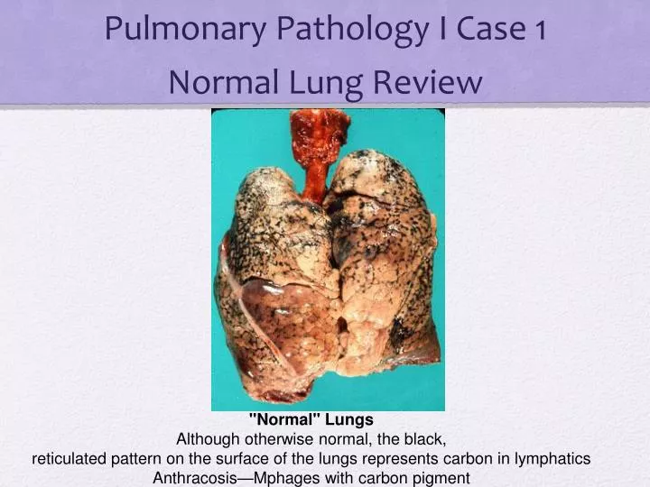
Pulmonary Pathology I Case 1 Normal Lung Review. "Normal" Lungs Although otherwise normal, the black, reticulated pattern on the surface of the lungs represents carbon in lymphatics Anthracosis—Mphages with carbon pigment. Pulmonary Pathology I, Case 2

E N D
Pulmonary Pathology I Case 1Normal Lung Review "Normal" Lungs Although otherwise normal, the black, reticulated pattern on the surface of the lungs represents carbon in lymphatics Anthracosis—Mphages with carbon pigment
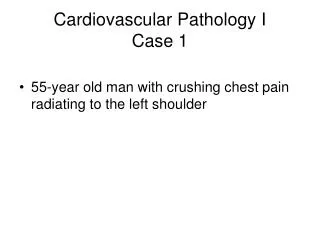
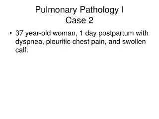
Pulmonary Pathology I, Case 2 Case 2/ 37 year-old woman, 1 day postpartum with dyspnea, pleuritic chest pain, and swollen calf • A 37-year-old woman develops dyspnea and pleuritic chest pain 1 day post-partum. She has a 20 pack-year history of smoking, which she gave up at the beginning of her pregnancy. On physical exam she has a fever of 38°C and a pulse of 102. Her lungs sound clear to auscultation. Her heart exam is normal, aside from tachycardia. Her left calf is swollen and tender. On chest x-ray a peripheral opacity is seen in the right lower lobe. • **You would have to prove that this is not a pulmonary embolism b/c of calf swollen & tender; post-partum (hypercoagulable state); pleuritic chest pain. • Peripheral Opacity if PE is a hemorrhagic infarct; Peripheral Opacity if Pneumonia is abscess! • Note that most PE’s have a normal CXR! You diagnose PE with V/Q scan or CT angiogram. • People can have PE’s but have no physical exam findings or symptoms!
a. Identify organ: lung; . Diagnosis: Pulmonary Embolism • b. Describe the characteristic pathologic changes in the histologic and gross specimen. • Low powerlots of blood—see RBC’s in the organ itself (“fill alveoli”)=hemorrhage • Closer updo see—very FULL vessels=congestion • Septal walls are thick and you don’t see nuclear detail of pneumocytes • (ischemic hemorrhagic necrosis—coagulative refers to the “ghost” like sxs) • Why is it hemorrhagic? • Dual blood supply—the bronchiolar vasculature feeds blood into the lung • WBC infiltration • d. What are risk factors for the development of this condition? • 95% of emboli arise from thrombi within large deep veins of lower legs (femoral) • Could also be from iliac, or endocarditis (throw clot from valve) • Virchow’s triad: Hypercoagulability, Stasis/abnormal blood flow, Endothelial injury • Watch bed rest, smokers, cancer, infections, CHF (stasis in heart), obesity, oral contraceptives, recent surgery (especially hip replacement & knee surgery) • e. What underlying genetic disorders may pre-dispose this individual to this problem? • Factor V leiden, prothrombin II gene mutation, Protein C, Protein S, or ATIII deficiency lupus anticoagulant • Treatment: UFH, LMWH’snote these do not bust the clot but prevent new formation
Case 2/ 37 year-old woman, 1 day postpartum with dyspnea, pleuritic chest pain, and swollen calf. Pulmonary Embolus The arrow points to an embolus extending through the branches of a pulmonary artery. Pulmonary Embolus/Infarct The embolus (arrow) lies in an opened pulmonary artery. The hemorrhagic infarct (*) is subpleural.
Case 2/ 37 year-old woman, 1 day postpartum with dyspnea, pleuritic chest pain, and swollen calf. Pulmonary Infarct Recent, small hemorrhagic pulmonary infarct Two/dual blood supplies: Pulmonary & Bronchial Note that infarcts are relatively uncommon
Case 2/ 37 year-old woman, 1 day postpartum with dyspnea, pleuritic chest pain, and swollen calf. Blood vessel partially occluded with organizing thrombus Hemorrhage RBC’s in tissue alveoli
Case 2/ 37 year-old woman, 1 day postpartum with dyspnea, pleuritic chest pain, and swollen calf. Note anthracotic areas (dark, are normal) and WBC infiltration (pathology) Alveoli with barely distinguishable (necrotic) septa Intra-alveolar hemorrhage
Saddle thrombus (arrow) is a blood clot which is seen at the bifurcation of the pulmonary artery. A saddle thrombus often results in sudden death with no pathologic change in the lungs
Case 3: 63 y/o man w/dyspnea, smokes 2 pack/day, hyperresonance of chest by percussion A 63-year-old man presents with dyspnea. He also complains of cough, which is worse in the mornings, off and on for several years. There is no history of fever or purulent sputum or cyanosis. He smokes two packs of cigarettes/day for the last 50 years. On exam there is hyper-resonance of the chest by percussion and prolonged expiratory phase on auscultation. Overall Case Analysis: Hyperresonant & prolonged expiratory phase—we’re thinking emphysema Cough, off and on—can be a component of chronic bronchitis 100 pack year history—major risk factor for COPD!! On XRAY: see blebs which are balls of air
a. Identify organ: lung/ Diagnosis: COPD: Emphysema • b. Describe the characteristic pathologic changes in the specimen. • See club-shaped ends to septa in emphysema • We would not be surprised to see squamous metaplasia (normal is columnar) • Destruction of septal wall of alveoli • Lots of black pigment in the septa • c. Correlate the clinical findings with the pathology. • Dyspnea—loss of functioning pulmonary parenchyma • Prolonged expiration: attempt to squeeze air out of lung • Purse-lip breathing—act to slow the respiratory rate and therby reducing dyspnea • Cough=chronic bronchitis • e. What is the most common etiology of this disorder? Tobacco/Smoking • When do they become symptomatic? Can be as early as 30’s or 40’s or when pts are older 50’s-60’s • f. What underlying genetic disorder(s) can contribute to this disorder? • Centroacinar emphysema=smoking • Panacinar emphysema=homozygous alpha 1 antitrypsin deficiency PiZZ phenotype (autosomal recessive)
Case 3: 63 y/o man w/dyspnea, smokes 2 pack/day, hyperresonance of chest by percussion Lungs and heart removed enbloc during autopsy See multiple blebs-balloons—they can pop and cause pneumothorax Emphysema Note the clusters of dilated air spaces which are conspicuous in the middle and lower lobes of the right lung and the lower lobe of the left lung. Both lungs are markedly enlarged.
Case 3: 63 y/o man w/dyspnea, smokes 2 pack/day, hyperresonance of chest by percussion Emphysema The cut surface of the lung reveals numerous dilated air spaces (alveoli). Cigarettes decrease antitrypsin action—so there less elastic activity of lung (because of increased elastases) (doesn’t bounce back, stays big); increase of compliance
Case 3: 63 y/o man w/dyspnea, smokes 2 pack/day, hyperresonance of chest by percussion Emphysema The cut surface of the lung reveals numerous (diffusely scattered) dilated air spaces.
Case 3: 63 y/o man w/dyspnea, smokes 2 pack/day, hyperresonance of chest by percussion Emphysema The photograph shows a large emphysematous, subpleural bleb.
Case 3: 63 y/o man w/dyspnea, smokes 2 pack/day, hyperresonance of chest by percussion Abnormally large alveoli; Could be a bleb
Case 3: 63 y/o man w/dyspnea, smokes 2 pack/day, hyperresonance of chest by percussion Large airspace caused by ruptured alveoli and alveolar septal destruction Septa with club-shaped end
Case 4: Pre-term infant (25 weeks) w/respiratory insufficiency Hyaline Membrane Disease--RDS • Case 4/A newborn develops respiratory insufficiency. He was born at 25 weeks gestation and weighed 500 gm at birth. The mother had fever for 3 days before delivery and the amniotic fluid was purulent. Surfactant (decrease surface tension) and antibiotics were administered. The baby’s condition deteriorated rapidly and he died 8 hours later. • a. Identify organ: lung • b. Describe the characteristic pathologic changes in the tissue. • on low power we don’t see air spaces, we see lots of blue, we see congested blood vessels • Hypercellularity makes the lungs very blue—which is normal for immature lung/stage of development—but is not ready for function—is atelectatic (missing surfactant) • Hyaline membrane formation occurs—protein and cellular debris • c. Diagnosis: • Note you can give them oxygen but oxygen can be injurious too!
Case 4: Pre-term infant (25 weeks) w/respiratory insufficiency Hyaline Membrane Disease The lungs are solid, airless and red-purple/ “beefy” (similar to the color of liver). These lungs are heavy and will sink in a cup of water (normal would float) On CXR—don’t see any black air spaces—only see whitish areas which indicate There is not air in the lungs/ “white out”. See “ground glass” picture (minimal air)
Case 4: Pre-term infant (25 weeks) w/respiratory insufficiency Hyaline membranes (protein, debris) Collapsed (atelectatic) alveoli No surfactant yet Immature lung
Case 4: Pre-term infant (25 weeks) w/respiratory insufficiency eosinophilic (pink) acellular hyaline membrane lining air spaces
Case 5: 44 y/o female getting chemo for ovarian cancer has sepsis ARDS: “Adult Respiratory Syndrome” or “DAD: Diffuse Alveolar Damage” • Case 5: A 44-year-old female undergoing chemotherapy for metastatic ovarian cancer is admitted to the hospital with a severe infection and sepsis. Over the next 2 days she develops progressive dyspnea. She becomes hypoxic and cyanotic and requires increasing oxygen concentration. Chest X-ray shows diffuse bilateral infiltrates. • a. Identify organ: lung--Diagnosis: ARDS • b. Describe the characteristic pathologic changes in the tissue. • Complement-driven process—macrophages, then NEUTROPHIL involvement, recruitment (chemotaxis) to the lung. • Neutrophils secrete all kinds of things that are destructive to type I pneumocytes, and type II pneumocytes will try to compensate and become hyperplastic • Septa are big because of FLUID; it is an edematous process because of the response to injury in lung • You don’t see fibrosis, you just see the “wispy” thickening of edema • You see hyaline membranes b/c of destruction of Type I pneumocytes, protein from exudation, organize into the membrane (does not have to do with surfactant)
Case 5: 44 y/o female getting chemo for ovarian cancer has sepsis/ARDS Adult Respiratory Distress Syndrome The lungs are large and dusky red (instead of pink and normal sized). The lungs are firm and airless by palpation. Would be 2 to 3 x heavier than normal lung On CXR: Normal size heart; diffuse “white out”; no pleural effusion (can see costophrenic angle)
Case 5: 44 y/o female getting chemo for ovarian cancer has sepsis Distended alveoli Greatly thickened septa (inflammation) Hyaline membranes Atelectatic alveoli
Case 5: 44 y/o female getting chemo for ovarian cancer has sepsis Hyaline membranes Intra-alveolar proteinacous debris, desquamated cells
Case 6: 56 y/o man with progressive dyspnea and bilateral infiltrates in lung Interstitial Fibrosis • Case 6/ A 56 year-old man develops progressive dyspnea over 6-8 months. He denies fever, chest pain or hemoptysis. He has never smoked cigarettes. On physical exam he is hypoxemic. On chest auscultation there are fine bi-basilar inspiratory crackles (sound like Velcro). Chest X-ray shows bilateral interstitial infiltrates with a peripheral distribution. • a. Identify organ: lung; diagnosis: • What would you do for this patient in your office? • Find out his occupational history—pneumoconoses? • Does he work more outside or on a farm now? • Does he have any new pets? Travel? New hobbies? ; PFTs to determine restrictive vs. obstructive and severity; Biopsy—open lung (because a needle aspiration is not going to help here) • b. Describe the characteristic pathologic changes in the tissue. • Fine Velcro crackles are classic for FIBROSIS • See lots of fibrosis in the lung, may see areas of immature collagen next to areas of mature collagen (would help us make diagnosis of IPF) • d. What pattern would be seen on this patient’s pulmonary function test? • Reduced lung complicance, reduced FVC
Case 6: 56 y/o man with progressive dyspnea and bilateral infiltrates in lung Idiopathic pulmonary fibrosis Cut surface of lung with fibrosis and anthracosis
Case 6: 56 y/o man with progressive dyspnea and bilateral infiltrates in lung Pulmonary fibrosis The cut surface of the lung reveals a "honeycomb" pattern of fibrosis. The large air spaces are accentuated by the thick walls of fibrous tissue.
Case 6: 56 y/o man with progressive dyspnea and bilateral infiltrates in lung Interstitial fibrosis Fibrous tissue replacing normal lung parenchyma Large airspaces separated by bands of fibrous tissue
Case 6: 56 y/o man with progressive dyspnea and bilateral infiltrates in lung Fibrosis Alveolus lined by reactive pneumocytes