Download
1 / 52
530 likes | 782 Views
Oral Mucosa III. Dr Jamal Naim PhD in Orthodontics. Nonkeratinocytes in the OE. It is a group of cells that differ from keratinocytes in having a clear halo around their nucleus. They are also termed clear cells ( They form a clear halo during histological processing)

E N D
Oral Mucosa III Dr Jamal Naim PhD in Orthodontics
Nonkeratinocytes in the OE • It is a group of cells that differ from keratinocytes in having a clear halo around their nucleus. • They are also termed clear cells (They form a clear halo during histological processing) • They represent a variety of cell types (10% of the cells in the oral Epithelium: • Melanocytes • Langerhans´ cells • Merkel cells • Lymphocytes
Nonkeratinocytes in the OE • They lack desmosomal attachments to adjacent cells • They have less number of tonofilaments than epithelial cells • They don’t participate in the process of maturation
Melanocytes • They are melanin producing pigment cells • They are situated in the stratum basale • They have an ectodermal origin • They divide and maintain themselves as a self-reproducing population • They have nor tonofilaments neither desmosomes • They posses dendritic processes that extend between keratinocytes
Melanocytes • They produce melanin in small structures called melanosomes • Melanosomes can be injected into keratinocytes by the dendritic processes
Oral pigmentation • The color of oral mucosa is the result of a number of factors: • Endogenous: melanin, hemoglobin etc. • Exogenous: foreign material induced into the oral epithelium such as amalgam • Lightly and darkly pigmented individuals have the same number of melanocytes, but their activity is different.
Langerhans cells • They are situated above the basal cell layer • They are also clear cells with dendritic processes • Unlike melanocytes they move in and out of the epithelium • Their source is the bone marrow • Their function is thought to be immunologic, that they recognize antigenic material and present it to T-lymphocytes
Merkel cells • They are situated in the stratum basale • They arise from division of keratinocytes • They haven't dendritic processes • Characteristic for the merkel cells are the membrane-bound vesicles. They liberate a transmitter substance across the junction between the merkel cell and adjacent nerve fibers • The function is thought to be sensitive to touch
Inflammatory cells • The most common one is lymphocyte • They are transient and don’t reproduce themselves in OE • They are often associated with Langerhans cells
Junction of the OE and lamina propria • The papillae of he connective tissue interdigitate with the epithelial ridges (rete pegs) • This arrangement makes the surface area of the interface larger than a simple flat junction and provide better attachment • Masticatory mucosa has the greatest number of papillae per unit area of mucosa • In lining mucosa the papillae are fewer and shorter
Basement membrane Connective tissue papillae
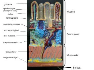
Lamina propria • It is the connective tissue of the oral epithelium • It consists of two layers: • The superficial papillary layer (associated with the epithelial ridges) • The reticular layer (reticular means netlike) • Both layers are different in the arrangement and concentration of collagen fibers
superficial papillary layer • reticular layer • Submucosa
Cells in the Lamina propria • Fibroblasts: • are the principal cells in the lamina propria • are responsible for turn over of fibers and maintenance of the tissue integrity • Macrophages • Mast cells • Inflammatory cells • Endothelial cells
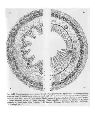
ORAL MUCOSA Oral epithelium Lamina propria Submucosa glands or fat cells Stratified squamous epithelium Papillary layer Reticular layer Keratinized May or may not be present Non- keratinized: • ortho-keratin. • para-keratin. • basal cell layer • Prickle cell layer • intermediate cell layer • superficial cell layer • Basal cell layer • Prickle cell layer • Granular cell layer • Cornified cell layer
Types of Oral mucosa • There are three main types of the oral mucosa, identified according their primary function: • Masticatory mucosa • Lining mucosa • Specialized mucosa • The larger part of the OM is lining mucosa (60%), followed by the masticatory (25%) and specialized mucosa (15%)
Oral Mucosa MASTICATORY SPECIALIZED DORSUM OF TONGUE GINGIVA, HARD PALATE LINING LOOSELY ATTACHED FIRMLY ATTACHED LIPS , CHEEKS, VENTRAL SURFACE OF TONGUE SOFT PALATE ALVEOLAR MUCOSA, FLOOR OF MOUTH VESTIBULAR FORNIX
Masticatory mucosa • Definition: Masticatory mucosa covers (25%) those areas that are exposed to compressive and shear forces and to abrasion during the mastication of food. • Hard palate • Gingiva • The epithelium of the masticatory is moderately thick and orthokeratinized.
Masticatory mucosa • Parakeratinization occur also in some areas • The junction between the OE and lamina propria is convoluted and the epithelial ridges are long • The lamina propria is thick with a dense network of collagen fibers in form bundles • It covers immobile structures and is bound firmly to them.
Masticatory mucosa • In some regions there is no submucosa, (mucoperiosteum) • In the lateral palatal regions the submucosa is thicker, interspersed with areas of fat and glandular tissue.\ • anterio-lateral (fatty) zone • Posterio-lateral (glandular) zone
palatine rugae incisive papilla antero-lateral (fatty) zone median palatine raphe postero-lateral (glandular) zone palatine gingiva soft palate uvula
Histology of the Hard Palate Epithelial ridges are long,irregular and numerous Mucosa Fatty zone Glandular zone Submucosa
Lining mucosa • It covers 60% of the oral cavity • The epithelium is thicker than in the masticatory mucosa and is nonkeratinized • The surface is flexible and able to withstand stretching • The junction between epithelium and connective tissue is smooth • The lamina propria is also thicker
LINING MUCOSA LOOSELY ATTACHED FIRMLY ATTACHED • LIPS • CHEEKS • VENTRAL SURFACE OF TONGUE, • SOFT PALATE • ALVEOLAR MUCOSA • VESTIBULAR FORNIX • FLOOR OF MOUTH
Loosely Attached “Movable”mucosa Alveolar mucosa loosely attached to periosteum Vestibular fornix allows mobility of lips and cheeks Floor of the mouth loosely attached to underlying structures allows mobility of the tongue
Firmly Attached Immovable mucosa Lip & cheek mucosa Inferior surface of the tongue Soft palate Oral side
ventral surface of the tongue Firmly attached to underlying muscles Thin non-Keratinized epithelium
Specialized mucosa • It covers the dorsal surface of the tongue • Although it is masticatory in function, it is also extensible • The mucosa of the tongue is composed of two parts covering: • The body of the tongue, the papillary part forms the anterior two thirds • The base of the tongue, the lymphatic part forms the posterior third.
Specialized mucosa • The body is derived from the second pharyngeal arch and the base from the third one. • The sulcus terminalis divides the tongue in the two parts
Specialized mucosa • The specialized mucosa contains different types of lingual papillae • Some of those papillae have a mechanical function, but others have a sensory function (taste) • The mucosa of the base of the tongue contains the lingual tonsils –extensive nodules of lymphoid tissue-
Lingual papillae • Fungiform papillae • Filiform papillae • Circumvallate papillae • Foliate papillae • Each type is differentiated to perform a specialized function while maintaining its primary role as a protective covering for the internal tissue components. All papillae contain a highly vascularized central connective tissue core (dermal papilla).
Fungiform papillae • Fungiform papillae resemble mushrooms in that they have a narrow stalk and a smooth-surfaced, dilated upper part. • These papillae, which contain scattered taste buds on their upper surfaces, which is Covered by thin Non-KE • They are irregularly interspersed among the filiform papillae. • They are located at the anterior portion of the tongue • They are red colored because of the high vascular connective tissue
Fungiform papillae • The papillae at the tip of the tongue are responsible for sweet taste and those at the lateral borders are responsible for salt sensation • Chorda tympani is responsible for these sensation salt sweet
Filiform papillae • They cover the entire anterior portion of the tongue • They are cone shaped structures, each with a core of connective tissue covered by a thick KE, characterized by curvature toward the back of the tongue. • Sometimes the filiform papillae are elongated through keratin buildups on their tip, so that the tongue become a hairy appearance, the hairy tongue
Filiform papillae • The mucosa between the filiform papillae is Non-KE and is highly extensible • They have a mechanical function, because they form an abrasive tough surface involved • in compression of food against • the palate • They don’t have taste buds
Circumvallate papillae • They are 8 to 12 circumvalate (walled) papillae located adjacent to the linea terminalis • They are large structures surrounded by a deep groove into which the open the minor salivary glands (the glands von Ebner) • They are composed of a connective tissue core and KE on the superior surface and Non-KE on the lateral walls • The Epithelium of the lateral walls contains taste buds
Circumvallate papillae • The Ebner glands are serous and are responsible for cleansing of the grooves and dissolving of the tastants • The circumvallate papillae are responsible for the sensation of bitter tastants by the glossopharyngeal nerve
Circumvallate papillae Taste buds
Foliate papillae • Foliate (leaflike) papillae are poorly developed in humans • Foliate papillae are located at the lateral margins of the posterior part of the tongue • They are composed of 4 to 11 parallel ridges that alternate with grooves • They contain some taste buds for sour sensation by the glossopharengeal nerve
Taste buds • Taste buds are specialized structures that contain the taste cells, the detectors of tastants. • They are located in: • All tongue papillae except the filiform ones • Soft palate • Posterior surface of the epiglottis • They are onion-shaped structures, each one containing 50–100 cells.
Taste buds • There are at least four qualities in human taste perception: saltiness, sourness, sweetness, and bitterness. • All qualities can be elicited from all the regions of the tongue that contain taste buds.