Download
1 / 35
350 likes | 454 Views
In this area of study, we are going to : Explore the various systems and mechanisms associated with the energy required for human movement. Consider the cardiovascular, respiratory and muscular systems and the roles of each in supplying oxygen and energy to the working muscles.

E N D
In this area of study, we are going to : • Explore the various systems and mechanisms associated with the energyrequired for human movement. • Consider the cardiovascular, respiratory and muscular systemsand the roles of each in supplying oxygen and energy to the working muscles. • Examine the way in which energy for activity is produced via the three energy systems and the associated fuels usedfor activities of varying intensity and duration. • Consider the many contributing factors to fatigue as well as recovery strategies used to return to pre-exercise conditions. • Participant in practical activities that explore the relationship between the energy systems during physical activity. • Complete One Test (20 marks) and one Laboratory based SAC (40 marks and 40 marks) AREA OF STUDY 2Physiological responses to physical activity
Key Knowledge • Mechanisms responsible for the acute responses to exercise in the cardiovascular, respiratory and muscular systems • Characteristics and interplay of the three energy systems (ATP – CP, anaerobic glycolysis, aerobic system) for physical activity, including rate of ATP production, the capacity of each energy system and the contribution of each energy system • Fuels (both chemical and food) required for resynthesis of ATP during physical activity and the utilisation of food for energy • Relative contribution of the energy systems and fuels used to produce ATP in relation to the exercise intensity, duration and type • Oxygen uptake at rest, during exercise and recovery, including oxygen deficit, steady state, and excess post-exercise oxygen consumption • The multi-factorial mechanisms (including fuel depletion, metabolic by-products and thermoregulation) associated with muscular fatigue as a result of varied exercise intensities and durations • Passive and active recovery methods to assist in returning the body to pre-exercise levels.
Key Vocabulary • Acute • Adenosine Diphosphate (ADD) • Plateau • Aerobic capacity • ATP-CP energy system • Reciprocal inhibition • Aerobic glycolysis • Cardiac Output • Respiratory Rate • Aerobic pathway • Chronic adaptations • Stroke Volume • Anaerobic capacity • Diffusion • Systolic Blood Pressure • Anaerobic glycolysis • Diastolic Blood Pressure • Tidal Volume • Anaerobic pathway • Heart Rate • Vasoconstriction • Anaerobic power • Lactate Inflection Point • Vasodilation • Arteriovenous Oxygen difference (a-vO2 diff) • Interplay • Ventilation • Adenosine Triphosphate (ATP) • Motor Unit • Ventilation Threshold • Phosphocreatine (PC)
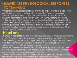
Acute Responses to Exercise • Immediate physiological responses to exercise are called ACUTE RESPONSES. • The body responds to the demands of exercise by making a number of physiological short-term changes to the cardiovascular, respiratory and muscular system. • Once exercise is stopped, these three systems will return to pre-exercise levels. THINKING TASK: BRAINSTORM ACUTE RESPONSES TO EACH SYSTEM
Acute Responses to Exercise Cardiovascular Muscular Respiratory Oxygen Consumption Temperature Ventilation A-V02 difference Motor Unit Recruitment Diffusion Cardiac Output Energy Substrates Venous Return Lactate Blood Pressure Redistribution of blood flow
Heart Rate Response to Exercise Laboratory Report Aim: To determine the change in heart rate in response to submaximal and maximal exercise. Equipment: Method: Subjects to sit quietly for 3-5 minutes. Record heart rate.
RESULTS • Input data into Excel and graph results DISCUSSIONG • Graph the heart rate responses for both submaximal and maximal exercise on the same axis. Describe the shape of each graph in terms of heart rate changes? • Did heart rate increase more with maximal exercise or submaximal exercise? Explain your answer with reference to physiological responses to exercise. • Why does heart rate need to increase with exercise? • Compare your data with that of another person n the class. Explain any differences that are evident. • Predict (by drawing on the graph) the heart rate response of a highly trained triathlete to this same activity. CONCLUSION Write a conclusion based on your results and the discussion.
Acute Respiratory Responses • Exercise places increased demands on the body’s need for oxygen to meet the rising energy demands of the activity • Ventilation increases prior to the begging of exercise and continues to rise to meet the oxygen demands of the exercise • Increases in ventilation are a result of an increase in tidal volume, respiratory rates or both • Gas exchange occurs at the alveolar-capillary interface (in lungs) and at the tissue-capillary interface (in the muscles) by process of diffusion
Respiratory Response to Exercise • Ventilation and Diffusion Read page 99-100 Complete Thinking Things Through (p.101)
Thursday 22nd March Thinking Things Through (101) 1. Respiratory rate and tidal volume. 2 Ventilation increases as the demand for oxygen increases and the need to remove carbon dioxide. Ventilation is increased through an increase in the respiratory rate (the number of breaths per minute) and/or the tidal volume (the amount of oxygen breathed in or out in one breath). Diffusion increases as a result of exercise. The increase in oxygen in the lungs causes an increase in diffusion of oxygen from the lungs into the blood and at the muscle, carbon dioxide levels are high so diffusion across the tissue-capillary interface increases also. 3 At rest the need for oxygen is low, but as exercise begins, the body’s need for oxygen increases significantly. To meet this need the body responds in a number of ways. The individual will breathe more often and more deeply (increased respiratory rate and increased tidal volume). These increases result in the rapid rise in ventilation as V = RR x TV. The levelling off represents a period of time where the oxygen demand is being met with supply (steady state) and no further increase in ventilation is required. The recovery section of the graph represents the gradual return of RR and TV to pre-exercise levels as the demand for oxygen has now decreased. 4 During submaximal exercise, the respiratory system will increase ventilation by increasing both TV and RR linearly, with respect to oxygen consumption, until a steady state is reached. At this point there will be no further increase in ventilation. During maximal exercise, ventilation will continue to increase until exercise ceases. The increase in ventilation is a result of increases in RR only. The rate of increase is linear up to the ventilator threshold, at which point ventilation
Acute Cardiovascular Responses to Exercise • During exercise, the CV system needs to deliver greater demands of oxygen and energy substrates to the working muscles • The focus is on getting more blood to the muscles to meet the increased oxygen demands and to speed up the removal of carbon dioxide and other waste products. TO DO THIS THE CARDIOVASCULAR SYSTEM UNDERGOES A NUMBER OF CHANGES
CARDIAC OUTPUT • Cardiac Output (Q): The amount of blood pumped by the heart in one minute. • Stoke Volume: is the amount of blood ejected by the left ventricle per beat. • Heart Rate is the number of times the heat beats in one minute • Cardiac Output is the product of stroke volume (SV) and Heart Rate (HR) Q= HR x SV • Increase in cardiac output increases blood pressure (BP) Explanation of Cardiac Output Read Table 4.2 and Figure 4.3 on Page 102 Copy these into your books
Copy Figure 4.4 (p.104) Describe what this graph is displaying BLOOD PRESSURE • During exercise, the increase in cardiac output (Q) results in an increased in blood pressure. • Using large muscles groups affects Systolic Blood Pressure more then diastolic.
VENOUS RETURN • As the cardiac output increases, it’s important that the venous return also increases. • The process of assisting venous return is done by one of 3 mechanisms • The muscle pump • The respiratory pump • Venoconstriction Copy Figure 4.6 (page 104) into books Read paragraph on page 105 to summarise the three mechanisms listed above.
Venous Return Describe the three mechanisms that allows venous return to occur? P. 104
Oxygen Consumption • Oxygen consumption is the volume of oxygen that can be taken up and used by the body. • As intensity of exercise increases, so does oxygen consumption. • This is a direct result of: • An increase in Cardiac Output (Q) • An increase in a-VO2 difference
Arteriovenous oxygen difference (a-VO2 Difference) • Difference in oxygen concentration in the arteries compared to the venuoles. • At rest, the arterial blood releases as little as 25% of it oxygen content to the tissue and the remaining 75 % is returned to the heart in the venous blood. • During exercise, the working muscles extract greater amounts of oxygen from the blood, increasing the a-VO2 difference. • While there is always some oxygen remaining in the blood returning to the heart, oxygen extraction can approach 100%. Copy Figure 4.8 (p.106) into books, with heading and a brief explanation.
Redistribution of Blood Flow • During exercise blood flow is redirected away from the spleen, kidneys, gastrointestinal tract and inactive muscles to the working muscles. • This is done so that the working muscles receive the greatest percentage of the cardiac output. • Copy Figure 4.7 (p.106) into books, with heading and a labels. • This process is assisted by vasoconstriction in the arterioles supplying the inactive areas of the body and by vasodilation in the arterioles supplying the working muscles. Explain the relationship between body temperature and redistribution of blood flow in the body as a result of continuous exercise.
Relationship between blood flow and temperature. • At submaximal exercise intensities blood flow is directed to the working muscles and to the skin to aid in temperature control. At maximal exercise intensities the increased demand for oxygen means that more blood is directed to the muscles and less to the skin (2 per cent) which means that temperature increases and the risk of heat related injuries increases.
Blood Volume • During exercise, blood volume decreases. • Plasma can decrease by 10% during prolonged exercise. Plasma decreases rapidly in the first 5 minutes of exercise, but then stablises. • The size of the decrease depends on intensity, duration and environmental factors (temperature, humidity, etc.) and level of hydration of the individual.
Acute Muscular Response to Exercise • When exercise commences, there is an increase in the rate of metabolism required to produce ATP. • This results in heat as chemical energy (fuel) is converted to mechanical energy (movement) • This causes the body’s temperature to increase • The body accommodates for these changes through increased blood flow, motor unit recruitment, using different energy substrates to fuel the body and remove lactate from working muscles
Motor Unit Recruitment • The motor unit is the means by which the central nervous system ‘talks’ to the muscles in order to control muscular contractions.
During exercise, the amount of force developed in the working muscles increases. To do this the brain • Increases the number of motor units recruited. • Or increases the frequency of messages sent to activate the motor units. If motor units always contact maximally, explain how the body controls movements that require more or less force?
Motor Unit Recruitment and Movement Fewer motor units are recruited for activities that require less force; more motor units are recruited for activities that require more force.
Energy Substrates • When exercise commences Adenosine Triphosphate (ATP) is the immediate energy source • However, this ATP is in short supply and when used up the muscles must rely on other energy substrates to fuel the body • Glycogen (stored energy) is used in both anaerobic and aerobic respiration to produce ATP. • During exercise phosphcreatine (PC) donates a phosphate to adenosine diphosphate (ADP) to resynthesis ATP. • The net result of exercise is a decrease in all fuel (ATP, PC, muscle glycogen) levels within the muscle. Study figure 4.3 )p.108). Copy the two diagrams into book and discuss. Why does the endurance athlete experience greater depletion in glycogen content than high-intensity sprint athletes?
Energy Substrate levels of a 100 metre sprinter and a marathon at the end of their event. marathon runner: decreased glycogen and intramuscular fat stores 100-metre sprinter: decreases ATP and CP stores
Lactate • As exercise starts large amounts of lactate are released from the muscles due to anaerobic production of ATP (without oxygen). • This means that during submaximal exercise there is a sharp increase in lactate • Lactate levels will rise until oxygen consumption can increase to meet energy demands of the muscle, and the lactate can be delivered to sites for removal. Lactate is present at rest and during submaximal and maximal exercise. However, it accumulates only at high exercise intensities. Discuss.
The Accumulation of Lactate At rest and during submaximal exercise intensities, lactate is produced, but sufficient oxygen is available for it to be broken down and removed by the body. At high intensities, lactate is being produced at higher rates than the body can clear it so it accumulates.
Acute Muscular Responses to Exercise • There are a number of mechanisms responsible for acute responses in the muscles • Increased blood flow -through redistribution there is more blood flowing to the muscles, delivery of large volumes of blood, increase to the surface area to increase diffusion rates • Increase in number of motor units recruited (dependent on the speed and strength required) • Decrease in Energy Substrates- ATP immediate source of energy, glycogen, phosphocreatine donation to resynthesis ADD to form APT. • Release of Lactate due to anaerobic production of ATP • Increase in heat production as a by-product of converting chemical fuel to movement energy. This increases core body temperature. The body must then employ methods ad mechanism to cool the body and restore homeostasis.
Acute Response to ExerciseLaboratory • Read Lab P. 112 • Before class, write aim, equipment, method and an empty results table (Respiratory Rate no Tidal Volume • Agree upon team sport to play (e.g. soccer, netball, basketball) • We will discuss the question in the last 15 minutes of class in the fitness centre.
AMAZING REVISION SITE! REVISION • prezi.com/msbfrxwphypl/acute-responses-to-exercise/ Links • Aerobic System (Aerobic glycolysis) • Fatigue-Fuel Depletion
Thinking things through p. 107 • 1 Cardiac output (Q) = stroke volume (SV) x heart rate (HR). Any increase in SV, HR or both will result in an increase in cardiac output. • 2 Resistance exercises cause compression of the blood vessels by the muscles causing an increase in blood pressure. Blood pressure can also increase due to the Valsalva response elicited in heavy resistance training, where air is forcefully expired against a closed airway. • 3 During exercise blood is redirected to the working muscles. This means more blood is delivered to the muscles and the muscles can extract greater amounts of oxygen to be used for energy production, causing an increase in a-vO2 difference. • 4 Each of the mechanisms has an impact on the others. Increases in ventilation and diffusion mean that more oxygen is available in the blood. Increases in cardiac output mean more blood is pumped out with each beat and delivered to the working muscles. The increase in venous return means that more blood is available to be ejected with each beat. Increases in cardiac output and a-vO2 difference lead to an increase in oxygen