Download
1 / 29
310 likes | 676 Views
Outline. vWFStructureLocationFunctionvWDHistoryClinical manifestations CategoriesDiagnosisTreatment. vWD. Family of bleeding disorders Caused by a deficiency or an abnormality of von Willebrand Factor. vWF. VWF gene : short arm of chromosome 12VWF gene is expressed in endothelial cells an

E N D
1. von Willebrand�s Disease December 3, 2004
2. Outline vWF
Structure
Location
Function
vWD
History
Clinical manifestations
Categories
Diagnosis
Treatment
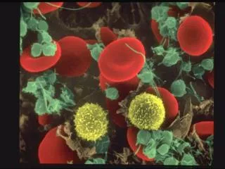
3. vWD Family of bleeding disorders
Caused by a deficiency or an abnormality of von Willebrand Factor
4. vWF VWF gene : short arm of chromosome 12
VWF gene is expressed in endothelial cells and megakaryocytes
vWF is produced as a propeptide which is extensively modified to produce mature vWF
Two vWF monomers bind through disulfide bonds to form dimers
Multiple dimers combine to form vWF multimers
5. vWF Production Vascular endothelial cells
Megakaryocytes
Most vWF is secreted
Some vWF is stored
Weibel-Palade bodies in endothelial cells
Alpha granules of platelets
Constitutive and stimulus-induced pathways
Release stimuli (EC)
Thrombin
Histamine
Fibrin
C5b-9 (complement membrane attack complex)
Release stimuli (platelets)
Thrombin
ADP
Collagen In plasma, the predominant MW is b/w 500,000-20,000,000
The bigger the multimer: more platelet and collagen binding sites more hemostatically competent
In plasma, the predominant MW is b/w 500,000-20,000,000
The bigger the multimer: more platelet and collagen binding sites more hemostatically competent
6. vWF Function Adhesion
Mediates the adhesion of platelets to sites of vascular injury (subendothelium)
Links exposed collagen to platelets
Mediates platelet to platelet interaction
Binds GPIb and GPIIb-IIIa on activated platelets
Stabilizes the hemostatic plug against shear forces
7. vW Factor Functions in Hemostasis Carrier protein for Factor VIII (FVIII)
Protects FVIII from proteolytic degradation
Localizes FVIII to the site of vascular injury
Hemophilia A: absence of FVIII
8. vWD History 1931: Erik von Willebrand described novel bleeding disorder
Hereditary pseudohemophilia
Prolonged BT and normal platelet count
Mucosal bleeding
Both sexes affected 1950s: Prolonged BT associated with reduced FVIII
1970s: Discovery of vWF
1980s: vWF gene cloned
9. Frequency Most frequent inherited bleeding disorder
Estimated that 1% of the population has vWD
Very wide range of clinical manifestations
Clinically significant vWD : 125 persons per million population
Severe disease is found in approximately 0.5-5 persons per million population
Autosomal inheritance pattern
Males and females are affected equally
10. vWD Classification Disease is due to either a quantitative deficiency of vWF or to functional deficiencies of vWF
Due to vWF role as carrier protein for FVIII, inadequate amount of vWF or improperly functioning vWF can lead to a resultant decrease in the available amount of FVIII
11. vWD Classification 3 major subclasses
Type I: Partial quantitative deficiency of vWF
Mild-moderate disease
70%
Type II: Qualitative deficiency of vWF
Mild to moderate disease
25%
Type III: Total or near total deficiency of vWF
Severe disease
5%
Additional subclass
Acquired vWD
12. Clinical Manifestations Most with the disease have few or no symptoms
For most with symptoms, it is a mild manageable bleeding disorder with clinically severe hemorrhage only with trauma or surgery Types II and III: Bleeding episodes may be severe and potentially life threatening
Disease may be more pronounced in females because of menorrhagia
Bleeding often exacerbated by the ingestion of aspirin
Severity of symptoms tends to decrease with age due to increasing amounts of vWF
13. Clinical Manifestations Epistaxis 60%
Easy bruising / hematomas 40%
Menorrhagia 35%
Gingival bleeding 35%
GI bleeding 10%
Dental extractions 50%
Trauma/wounds 35%
Post-partum 25%
Post-operative 20%
14. vWD Type I Mild to moderate disease
Mild quantitative deficiency of vWF
vWF is functionally normal
Usually autosomal dominant
Penetrance may vary dramatically in a single family
15. vWD Type 2 Usually autosomal dominant
Type 2A
Lack high and intermediate molecular weight multimers
Type 2B
Multimers bind platelets excessively
Increased clearance of platelets from the circulation
Lack high molecular weight multimers Type 2C
Recessive
High molecular weight vWF multimers is reduced
Individual multimers are qualitatively abnormal
Type 2M
Decreased vWF activity
vWF antigen, FVIII, and multimer analysis are found to be within reference range
Type 2N
Markedly decreased affinity of vWF for FVIII
Results in FVIII levels reduced to usually around 5% of the reference range.
16. vWD Type III Recessive disorder
vWF protein is virtually undetectable
Absence of vWF causes a secondary deficiency of FVIII and a subsequent severe combined defect in blood clotting and platelet adhesion
17. Acquired vWD First described in 1970's
fewer than 300 cases reported
Usually encountered in adults with no personal or family bleeding history
Laboratory work-up most consistent with Type II vWD
Mechanisms
Autoantibodies to vWF
Absorption of HMW vWF multimers to tumors and activated cells
Increased proteolysis of vWF
Defective synthesis and release of vWF from cellular compartments
Myeloproliferative disorders, lymphoproliferative disorders, monoclonal gammopathies, CVD, and following certain infections
18. vWD Screening PT
aPTT
(Bleeding time)
19. vWD: aPTT and PT aPTT
Mildly prolonged in approximately 50% of patients with vWD
Normal PTT does not rule out vWD
Prolongation is secondary to low levels of FVIII
PT
Usually within reference ranges
Prolongations of both the PT and the aPTT signal a problem with acquisition of a proper specimen or a disorder other than or in addition to vWD
20. vWD and Bleeding Time Historically, bleeding time is a test used to help diagnose vWD
Lacks sensitivity and specificity
Subject to wide variation
Not currently recommended for making the diagnosis of vWD
21. vWD Diagnostic Difficulties vWF levels vary greatly
Physiologic stress
Estrogens
Vasopressin
Growth hormone
Adrenergic stimuli
vWF levels may be normal intermittently in patients with vWD
Measurements should be repeated to confirm abnormal results
Repeating tests at intervals of more than 2 weeks is advisable to confirm or definitively exclude the diagnosis, optimally at a time remote from hemorrhagic events, pregnancy, infections, and strenuous exercise
vWF levels vary with blood type
22. vWD Diagnosis Ristocetin
Good for evaluating vWF function,
Results are difficult to standardize
Method
Induces vWF binding to GP1b on platelets
Ristocetin co-factor activity: measures agglutination of metabolically inactive platelets
RIPA: metabolically active platelets
Aggregometer is used to measure the rate of aggregation
vWF Antigen
Quantitative immunoassay or an ELISA using an antibody to vWF
Discrepancy between the vWF:Ag value and RCoF activity suggests a qualitative defect
Should be further investigated by characterization of the vWF multimeric distribution
23. Additional Assays Multimer analysis
PFA-100 closure time
Screens platelet function in whole blood
Prolonged in vWD, except Type 2N
FVIII activity assay
24. vWD Treatment DDAVP
Cryoprecipitate
FVIII concentrate
25. vWD and DDAVP Treatment of choice for vWD type I
Synthetic analogue of the antidiuretic hormone vasopressin
Maximal rise of vWF and FVIII is observed in 30-60 minutes
Typical maximal rise is 2- to 4-fold for vWF and 3- to 6-fold for FVIII
Hemostatic levels of both factors are usually maintained for at least 6 hours
Effective for some forms of Type 2 vWD
May cause thrombocytopenia in Type 2b
Ineffective for vWD Type 3
26. Factor VIII Concentrates Alphanate and Humate P
Concentrates are purified to reduce the risk of blood-borne disease
Contain a near-normal complement of high molecular weight vWF multimers
27. vWD Treatment Platelet transfusions
May be helpful with vWD refractory to other therapies
Cryoprecipitate
Fraction of human plasma
Contains both FVIII and vWF
Medical and Scientific Advisory council of the National Hemophilia Foundation no longer recommends this treatment method due to its associated risks of infection
FFP
An additional drawback of fresh frozen plasma is the large infusion volume required
28. References Castaman G, et al. Haematologica, 88(01):January 2003
Harmening, Denise. Clinical Hematology and Fundamentals of Hemostasis. 1997.
http://www3.ncbi.nlm.nih.gov/entrez/dispomim.cgi?id=193400