Download
1 / 58
580 likes | 780 Views
EKG ROUNDS Bundle Branch Blocks. 07. 04. 2005 Nadim Lalani. OUTLINE. RBBB LBBB HEMiBLOCKS/ FaSCicULAR BLOCKS. Case. A 13-year-old male.presents with a history of Syncpoe: What else do you want to know on hx?. Brugada Syndrome:. Described in 1992:

E N D
EKG ROUNDSBundle Branch Blocks 07. 04. 2005 Nadim Lalani
OUTLINE • RBBB • LBBB • HEMiBLOCKS/ FaSCicULAR BLOCKS
Case • A 13-year-old male.presents with a history of Syncpoe: • What else do you want to know on hx?
Brugada Syndrome: • Described in 1992: • Syndrome consisting of syncopal episodes and/or sudden death in patients with a structurally normal heart. • Characteristic ECG with a pattern of right bundle branch block with an ST segment elevation in leads V1 to V3 was • In 1998 recognised poor prognosis of patients with the syndrome not receiving an implantable defibrillator.
Brugada Syndrome: • Epid: incidence of 0.05% to 0.6%. Mostly SE Asian / Japan an autosomal dominant mode of transmission. • Who?: Adults 30-40 (first case reports were kids) • Related to defect of SCN5a Fast sodium channel predisposing pt to re-entry circuit.
Brugada Syndrome: Clinical manifestations • The complete syndrome is characterised by episodes of rapid polymorphic VT in patients. Resulting in syncopal attacks. • When the episodes are sustained, cardiac arrest and eventually sudden death occur. • There exist asymptomatic individuals in whom the atypical ECG is detected during routine examination. • NB now known some variation in the EKG (three types of ST changes/ a-fib &c)
Brugada Syndrome: • Diagnosis: • the typical ECG pattern and there is a history of aborted sudden death or syncopes caused by a polymorphic VT. • There are also many patients with a normal ECG. Syndrome only recognised a posteriori when the typical pattern appears in a follow-up ECG or after the administration of pro-arrythmics. • Additional diagnostic problems are caused by the changes in the ECG induced by the autonomous system and by antiarrhythmic drugs.
Brugada Syndrome: • Prognosis and treatment: implantation of a cardioverter-defibrillator is mandatory in these patients. Case report of a 23 yold patient being managed on amiodarone until implantation
Case • Mrs M. 39 yo female had a pulmonary artery prosthesis and ASD repair 3 weeks ago. Helathy otherwise . On Lipitor. Presents with migratory arthralgias/myalgias. • EKG Diagnosis? • Axis?
RBBB • Three phases: Septal depolarisation. Left ventricular depolarization. Delayed stimulation of the right ventricle.
RBBB septal depolarization produces a small septal r wave in V1 and a small septal q wave in V6 depolarization of LV produces an S in V1 and an R in V6 delayed right ventricular depolarization produces a wide R wave in V1 and a wide S wave in V6
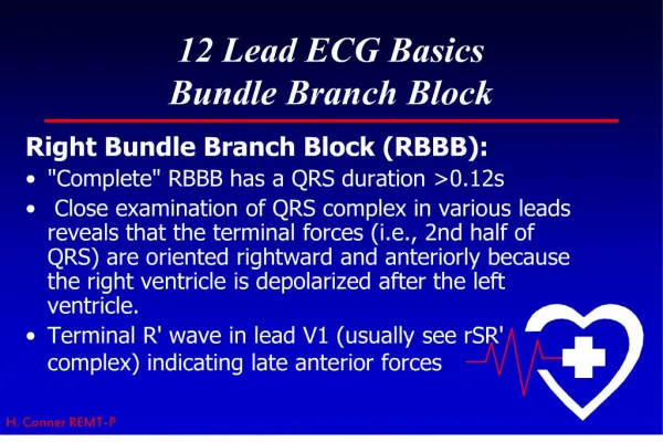
RBBB Summary • Right Leads ( V1 eg) show an rSR’ complex with a wide R wave. • Occasionally, however, the S wave never quite makes its way below the baseline. Consequently, the complex in lead V1 has the appearance of a large notched R wave (“rabbit ears”) • Left Leads (I,V6) show a qRS pattern with a wide S wave.
Case 2 • Mr R . 55 yo admited to FMC after head-on MVC in which he sustained Multiple rib #s,# L Hip and # L ulna. 2 days post-admit develops acute SOB with sat 80%. • EKG Diagnosis? • Axis? • Comment on T waves?
RBBB, ?RAD, TWI’s Primary TWI’s
ST changes • TWI’s in the right chest leads are a characteristic finding with RBBB. • These inversions are referred to as secondary changes because they reflect just the delay in ventricular stimulation. • By contrast, TWI’s in V5,V6 are primary T wave abnormalities reflect an actual change in repolarization (ie ischemia &c.)
Case 3 • 80 yo guy with HTN. Presents with this EKG:
COMPLETE AND INCOMPLETE RBBB • Depends on the width of the QRS complex: Complete RBBB is defined by a QRS that is 0.12 second or more with an rSR in lead V1 and a qRS in lead V6. Incomplete RBBB shows the same QRS patterns, but its duration is between 0.1 and 0.12 second
Case 4: • 77 yo M with HTN, and known CAD:
Left Bundle Branch Block • With LBBB the septum depolarizes from right to left .Therefore the EKG loses the normal septal r wave in lead V1 and the normal septal q wave in lead V6. • Left ventricular depolarization is prolonged, yielding a wide QRS. • Lead V6 you see a wide, entirely positive (R) wave. • In the right chest leads (e.g., V1 ) you see a negative QRS (QS) complex.
Lose septal r in V1 And septal q in V6 Wide QS (sometimes rS) in V1 Wide R in Left Leads
Congenital septal lesions, CAD, anterior MI (occlusion of proximal LAD), pulmonary hypertension, normal variant in 0.2% adults
R’ R r q S S R QS
Fascicular Blocks (Hemiblocks) • The RBB is a single pathway consisting of just one main fascicle or bundle. • In contrast the LBB has an anterior fascicle and a posteriorfascicle.
EKG Changes: • Hemiblock (unlike a full LBBB or RBBB) does not widen the QRS complex. • Main effect is a change in the QRS axis: Left anterior fascicular block results in marked left axis deviation (-30° or more) Left posterior fascicular block produces marked right axis deviation (+120° or more).
Left anterior hemiblock shifts the QRS axis to the left by delaying activation of the more superior and leftward portions of the left ventricle. • Left posterior hemiblock shifts it to the right by delaying activation of the more inferior and rightward portions of the left ventricle. • In both cases the QRS axis therefore is shifted toward the direction of delayed conduction.
Axis? Between -30 and -90 deg LAD
Axis? RAD
Case • 67 yo M Obese, Hx CHF. Presents with SOB: • EKG Dx? • Is he having an MI?
Sgarbossa et. al. : • 131 patients from GUSTO-I trial who had LBBB with MI. Matched with controls. Came up with following criteria: ST-segment elevation . 1 mm concordant with QRS complex – 73 % sens and 92 % spec for acute MI. ST-segment depression . 1 mm in lead V1, V2 or V3 – highly specific (96%) but less sensitive (25%) for MI ST-segment elevation . 5 mm discordant with QRS complex – 31 % sens, 92 % spec.
Concordant ST ↑ ST ↓ Discordant ST ↑ ST ↓
Shlipak MG, Lyons WL, Go AS, et al. Should the electrocardiogram be used to guide therapy for patients with left bundle branch block and suspected myocardial infarction? JAMA 1999;281:714–19.: • Reviewed patients presenting with LBBB and an acute cardiopulmonary history and assessed the usefulness of the Sgarbossa criteria. • Criteria had a sensitivity of 10% and a specificity of 100%. • Most (90%) patients with AMI will not meet the criteria. • Support thrombolysing all patients (except those with contraindications) who have a history suggestive of AMI and LBBB.
Case A • 65 yo M smoker with DM presents with 4 hours RSCP: • EKG Diagnosis? • Axis?
Case B RBBB, ? Primary TWI’s V4,V5
Case C LBBB