Download
1 / 47
500 likes | 564 Views
Tolerance and autoimmunity. Tolerance = specific unresponsiveness to a specific antigen „ Horror autotoxicus ” Paul Erlich Central tolerance – unresponsiveness of the developing immature lymphocytes in the central lymphoid organs against self

E N D
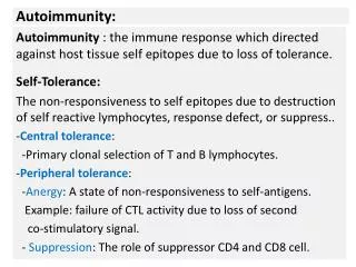
Tolerance and autoimmunity • Tolerance = specificunresponsivenessto a specificantigen • „Horror autotoxicus” Paul Erlich • Centraltolerance– unresponsiveness of thedevelopingimmaturelymphocytesinthecentrallymphoidorgansagainstself • Peripheraltolerance– maturelymphocytesintheperiphery, dependsonthewayencounteringantigen • Antigens that induce tolerance are called tolerogens • Whatmechanismsplayingroleintolerance? • Clonaldeletion • Receptor editing • Anergy (largeantigendose, longtimestimuli) • Clonalignorance (smalldoseantigen, lowaffinitybinding) • Immunologicalsuppression (suppressorcellsorfactors)
Autoimmunity and tolerance • autoimmune disease : immune response against the seemingly healthy self structure • lack of tolerance, disturbed regulation • the regulated autoreactivity is physiological (autoantibodies and autoreactive cells are present in health) • tolerance: regulated unresponsiveness to antigens for which the they should respond (clonal deletion, anergy, suppression) • Clonal deletion (T cells), inactivation (anergia) (B cells), or the results of active immunesuppresion
Four cellular strategies are used to regulate self-reactive receptors at different points during B- and T-cell differentiation
A map of the cellular checkpoints regulating self-reactivereceptors (19) BCR-induced death of germinalcentreB cells; (20) Germinal-centre B-cell death from competition forfollicular T helper cells. (21) Controlof autoantibody accumulation and inflammation in tissues. (6) BCR tuning/anergy (8) Follicular exclusion of B cells; (9) B-cell competition forBAFF; (1) Arrest ofimmature B-cell maturation; (2) BCR light chain editing by V(D)Jrecombination; (3) Death and deletion of immature B cells. (11) Controls on availability of extrafollicular T-cell help; (12) Control of TLR ligands and signalling; (13) B-cell death induced byFASL from T cells (14) BCR inhibition of plasma-cell differentiation (10) T-cell competition for IL-7. (7) TCR tuning/anergy; (15) Control of B7 ligands and other costimulatory molecules; (16) T-celldeath induced by FASL; (17) T-cell suppression by TR cells. (4) TCR –chainediting by V(D)J recombination; (5) Death and deletion of semi-mature T cells
. . . B newborn A X A transplant from strain B remains, while from strain C is not Cells from strain B C Immunological tolerance Peter Medawar The Nobel Prize in Physiology and Medicine 1960 Immunological Tolerance Self = Immunological self! Tolerance
physical barrier • no lymph circulation • immunosuppressive factors (e.g. TGFb) • low MHC expression • FasL • Implants are not rejected, no immune response
Damage to an immunologically privileged site can induce an autoimmune response sympathetic ophthalmia
Central tolerance Periferal tolerance
Thymus • Positive selection: cells that are able to recognize and bind to self MHC or to peptide + MHC molecules are selected to grow • Negative selection: cells that recognize and efficiently bind self peptides are auto-reactive cells and undergo apoptotic cell death because they are harmful to the host • Cells that pass both positive and negative selection tests “graduate” from thymus ; enter circulation as mature T lymphocytes
Central T celltolerance. Recognition of self antigens by immature T cells in the thymus may lead to death of the cells (negative selection, or deletion) or the development of regulatory T cells that enter peripheral tissues.
The differentiation model of promiscuous gene expression in thymic epithelial cells. AIRE: autoimmune regulator APECED: AIRE mutation Autoimmune polyendocrinopathy-Candidiasis-Ectodermal distrophy
Diverse tissues are represented by promiscuous gene expression in medullary thymic epithelial cells (mTECs).
The role of Aire in immune tolerance. Aire drives the expression of certain organ-specific genes in medullary thymic epithelial cells (TECs). Immature T cells recognizing these self molecules are deleted from the repertoire. Mutations in the AIRE gene are the cause of a multiorgan autoimmune disease called the autoimmune polyendocrine syndrome (APS).
Models of the cellular source of IL-2 for Treg cells Thymus, secondary lymphoid organs Treg cells do not produce IL-2
Mechanisms of peripheral T cell tolerance. The signals involved in a normal immune response (A) and the three major mechanisms of peripheral T cell tolerance (B)
Mechanisms of T cell anergy If the T cell recognizes a self antigen without costimulation, the T cell becomes unresponsive to the antigen because of a block in signaling from the TCR complex or engagement of inhibitory receptors (such as CTLA-4). The signaling block may be the result of recruitment of phosphatases to the TCR complex or the activation of ubiquitin ligases that degrade signaling proteins. The T cell remains viable but is unable to respond to the self antigen.
Regulatory T cells. Regulatory T cells are generated by self antigen recognition in the thymus and (probably to a lesser extent) by antigen recognition in peripheral lymphoid organs. The development and survival of Treg requires IL-2 and the transcription factor FoxP3
Autoreactive B cellsdonotcompeteeffectivelyinperipherallymphoidtissueto enter primarylymphoidfollicles. Inthe top panel, B cellsareseen entering theT-cellzone of a lymphnodethroughhighendothelialvenules (HEVs). Thosewithreactivityforforeignantigensareshowninyellow; autoreactivecellsaregray. The autoreactivecellsfailtocompetewith B cellsspecificforforeignantigensforexitfromtheT-cellzone and entryintoprimaryfollicles (center panel). The autoreactive B cellsthatfailtoreceivesurvivalsignals most likelyundergoapoptosisin situintheT-cellzone (bottom panel).
Cell surface receptors Intracellular molecules Keio J Med 2004; 53 (3): 151–158
Regulation of B cell activation by inhibitory receptors Defectivesignalingmaybreaktolerance
Overall view of activatingFcreceptors and inhibitoryFcγ RIIB, and relatedautoimmunediseases
. Spontaneousautoimmunity FcgRIIb KO mice: C57BL/6BALB/c anti-DNA resistanttonuclearantigen glomerulonephritis susceptibilityloci: chr 12 és 17 FcRgKO: NZB/W F1: lupus nephritis ” FcRg -/-: IC deposition, Complementactivationbutless kidneyinjury ”FcgRIIb-/-: more seriousdisease FcRdefectsinNOD mice and EAE model
. . . . . Inducedautoimmunity Collagen induced arthritis: CIAC-II DBA/1 mice, FcRg-/- protected FcgRIIb -/- : enhancedanti-CIIIgG, CIA arthritis more C57BL/6 : afterFcgRIIdeletion : CIA Goodpasture’s szindróma: a-CII a-CII a-CII FcgRIIb -/- Pulmonaryhaemorhages Glomerulonephritis EnhancedantibodylevelC-IV collagen-IV: FcRgdeficientmice: no evidence of disease
FcgRpolymorphisminautoimmunediseases (human) AutoimmunediseaseFcgRIIaFcgRIIbFcgRIIIaFcgRIIIb -------------------------------------------------------------------------------------------------- SLE131 Arg - - - - - 158 Phe - - 232 Thr158 Phe - - - - NA2 RA - - 158 Phe - Wegener’s granulomatosis - - - NA1 Guillain-Barre syndroma131 Arg - - NA2 Multiple Sclerosis131 Arg - - NA2
B10 cell regulatory effects in autoimmune disease. Kalampokis et al. Arthritis Research & Therapy 2013.
Selfregulation of theimmuneresponse • elimination of antigen • inhibition of thespecificimmuneresponse • regulation of thetype and size of immuneresponse • Tolerance is a result of an activeprocessevokedbythetolerogen • Lack of regulation: autoimmunediseases
Bacterial adjuvants are required for induction of experimental autoimmune disease Experimentalallergicencephalomyelitis – EAE: model of sclerosis multiplex Mycobacterium tuberculosis in complete Freund adjuvant
Virus infection can break tolerance to a transgenic viral protein expressed in pancreatic B cells LCMV = lymphocytic choriomeningitis virus
There are several ways in which infectious agents could break self-tolerance
Amino acid changes in the sequence of an MHC class II protein correlate with susceptibility to and protection from diabetes Asp 57, shown in red on the backbone structure of the DQβ chain, forms a salt bridge (shown in green in the center panel) to an arginine residue (shown in pink) in the adjacent α chain (gray). The change to an uncharged residue (for example, alanine, shown in yellow in the bottom panel) disrupts this salt bridge, altering the stability of the DQ molecule Asp – Val, Ser, Ala - IDDM HLA-DQb1
Associations of HLA serotype and sex with susceptibility to autoimmune disease. SLE
Genes encoding three functional classes of proteins are implicated in the etiology of SLE
Autoimmune diseases classified by the mechanism of tissue damage
Autoantibodies inhibit receptor function in myasthenia gravis
Schematic representation of a local autoantibody-induced inflammatory network in an arthritic joint.
Possible mechanisms of the onset of autoimmune diseases: - disturbed regulation: CD8 , TH1 - TH2 - molecular mimicri: (Streptococcus M protein – myosin in heart reumatic feaver) crossreaction between self and microbes - new T cell epitopes appear: T cell bypass- escape from regulation xenogen proteins (RBC, MBP), virus-modified self - appearance of hidden structures: from damaged cells, tissues autoreactive antibodies - elimination, intracellular proteins quantities changed - MHC antigen presentation is modified (diabetes cells, Hashimoto tyroiditis) aberrant MHCII expression – modified by cytokines (IFN, TNF) - inflammatory cytokines: IL-1, IFN, TNF, increased antibody production Role of HLA: Allow or protect upon presentation of autoantigenes correlation between MHC genotype – susceptibility for autoimmune desease (e.g.:IDDM:DQ*0301 )