Download
1 / 59
590 likes | 595 Views
Recombinant DNA. OVERVIEW OF RECOMBINANT DNA TECHNOLOGY. Recombinant DNA technology allows a DNA fragment from any source to be joined in vitro with a nucleic acid vector that can replicate autonomously in microorganisms.

E N D
OVERVIEW OF RECOMBINANT DNA TECHNOLOGY • Recombinant DNA technology allows a DNA fragment from any source to be joined in vitro with a nucleic acid vector that can replicate autonomously in microorganisms. • This provides a means of analyzing and altering genes and proteins. It provides the reagents necessary for genetic testing for carrier detection and prenatal diagnosis of genetic diseases and for gene therapy. • Additionally, this technology can provide a source of a specific protein, such as recombinant human insulin, in almost unlimited quantities.
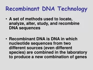
Two approaches to producing recombinant DNA for cloning have been developed for use with somewhat different applications: • Cloning restriction fragments of cellular DNA • Cloning cDNA produced by reverse transcription of cellular mRNA
Cloning Restriction Fragments of Cellular DNA • Cloning DNA restriction fragments is useful in the following applications: • Sequencing DNA (Human Genome Project, Genetic diagnosis) • Producing restriction maps for gene mapping • Studies involving non-expressed DNA sequences
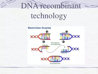
The first step in this procedure is to produce restriction fragments of DNA using restriction endonucleases. Restriction Endonucleases • These enzymes are isolated from bacteria, their natural source. • There are many different restriction endonucleases isolated from a variety of bacteria that are now readily available commercially. • In bacteria they act as part of a restriction/ modification system that protects the bacteria from infection by DNA viruses.
Restriction endonucleases recognize double-stranded DNA sequences called palindromes (inverted repeats) usually of four to eight base pairs in length. For example, Figure 1-6-1 shows the recognition site for EcoRI, a restriction endonuclease isolated from Escherichia coli. • A palindrome can be identified by examining the sequence of only one strand. Draw a line through the center of the sequence (through the central base for palindromes with an odd number of nucleotides). If the sequence is folded along this line the bases should pair.
Prokaryotic Restriction Modification Systems • Provide defense against infecting DNA viruses • Methylase enzyme modifies and protects palindromes in bacterial DNA. • Unmethylated palindromes of infecting viral DNA are recognized by restriction endonuclease. • Viral DNA is fragmented and destroyed.
DNA from a source to be cloned is mixed with a particular restriction endonuclease such as EcoRI, producing DNA restriction fragments. • Some restriction endonucleases such as EcoRI produce asymmetric cuts within the palindrome yielding "sticky ends" on the fragments. • Sticky ends are advantageous in facilitating the recombination of a restriction fragment with the vector DNA. • Others, like HaeIII, cut both strands in the same location yielding "blunt ends" on the restriction fragments.
Cloning Restriction Fragments Using Vectors • To clone the restriction fragments, they must each be inserted into a vector. • A vector is a piece of DNA (plasmid, viral chromosome, yeast chromosome) capable of autonomous replication in a host cell, for instance, plasmid pBR322. • The DNA used as a vector usually has: - At least one type of palindrome recognized by a restriction endonuclease - An origin for autonomous replication - At least one gene for resistance to an antibiotic
Cloning vectors • Common properties • origin of DNA replication • unique restriction sites for insertion of DNA • multiple cloning sites containing many restriction sites engineered into many plasmid vectors • Easy identification and recovery of clones • Types of vectors • plasmids containing drug resistance gene (up to 10 kb) • many commercially available plasmids • bacteriophage, e.g., lambda (~ 15-20 kb) • cosmids for larger DNA molecules (~ 40-50 kb) • BAC: bacterial artificial chromosome (~ 100-300 kb) • YAC: yeast artificial chromosome (~ 1000 kb)
The vector is cut with the restriction endonuclease and mixed with the DNA restriction fragments to be cloned. As shown in Figure 1-6-3, once the vectors have combined with one of the restriction fragments, DNA ligase is used to form permanent PDE bonds between the fragment and the vector. This produces recombinant DNA.
Once the recombinant vectors have been produced, they are used to transform host cells. • In the example of the plasmid pBR322, the host cells are bacteria. Once transformed, the bacteria are plated on selective media so that bacteria transformed with a recombinant plasmid can be easily identified. • In the case of plasmid pBR322, bacteria with recombinant plasmids would be resistant to ampicillin but sensitive to tetracycline.
The collection of colonies produced is referred to as a genomic DNA library. The library must be screened with a radioactive probe to identify the colony with the desired restriction fragment.
Uses of Genomic Libraries • Large quantities of each clone can be grown for DNA sequencing studies, similar to what is being done in the Human Genome Project. • By producing genomic libraries using different restriction endonucleases (or allowing one type of restriction endonuclease to digest a DNA sample for different times), regions of overlap can be identified and the fragments ordered, producing DNA restriction maps useful for genetic testing and sequencing. • Other genetic markers may be identified in this way, such as minisatellite and microsatellite sequences. • Genomic libraries are also useful to clone and study DNA sequences that are not expressed in cells (response elements, introns, promoters).
Restriction Maps • A restriction map is a linear (or circular) map of the order and distances of restriction endonuclease cut sites in a segment of DNA • Each DNA fragment, no matter what size, has its own unique restriction map • Restriction maps yield structural info. They are useful in comparing DNA fragments to look for regions of identity
Restriction Maps • Line drawings of DNA identifying sites cut by restriction endonucleases. • Identify potential RFLP markers for genetic diagnosis. • Example: Restriction site polymorphism for Mstll may be used to identify individuals with the sickle cell mutation
The difference between the standard BA allele and the sickle-cell BS allele is a single-nucleotide substitution (A>T) in the second position of the sixth codon of this gene. The sequence of the standard BA allele (CCTGAGG) happens to correspond to an MstII restriction site (CCTNAGG), which is altered in the BS allele (CCTGTGG). The beta-globin gene region includes two additional MstIIsites
Cloning cDNA Produced by Reverse Transcription of Cellular mRNA • If the end goal of cloning is to have a cloned gene expressed in a cell, the entire coding sequence must be cloned intact. Furthermore, if a cloned eukaryotic gene is to be expressed in bacteria (to make recombinant proteins), the gene must not contain introns, which could not be processed in a prokaryotic cell. In these cases it is more convenient to clone cDNA rather than DNA restriction fragments.
Producing cDNA by Reverse Transcription of mRNA • Cytoplasmic mRNA is isolated from a cell known to express the desired gene. Reverse transcriptase, along with other components (Figure 1-6-4), is used in vitro to produce double stranded cDNA that is subsequently recombined with a chosen vector to produce the recombinant DNA for cloning. In this approach: • All genes expressed will be cloned along with the desired gene. • None of the non-expressed DNA in the cell will be cloned*. • Each cDNA represents the complete coding sequence of a gene. • The cDNAs have no introns. • An expression library is produced at the end of the cloning procedure.
Expression Vectors • If the goal of the cloning procedure is to obtain a recombinant protein, appropriate sequences required for transcription and translation in the cloning host cell must be provided because they will not be part of the cDNA. • For instance, to produce recombinant human insulin in bacteria, a bacterial promoter and a Shine-Dalgarno sequence must be included in the cloning plasmid near the insertion site for the cDNA. • Figure 1-6-5 shows an example of an expression vector, pUC. In some expression vectors, other regulatory sequences such as operators are added to allow expression of the cloned gene to be controlled.
Uses of cDNA (Expression) Libraries • Once the recombinant expression vectors containing the cDNA inserts are produced, they are used to transform bacteria (or other host cells) and produce cDNA (expression) libraries. Expression libraries may be used to produce recombinant proteins that in some cases have significant advantages over isolating them from natural sources: • Larger quantities may be produced. Recombinant human insulin used to treat diabetics. • Natural source may carry risk of infection. Recombinant Factor VIII used to treat hemophilia A has helped reduce the incidence of HIV infection in hemophiliacs. Recombinant HbsAg is now used to immunize against hepatitis B, eliminating the risk of introducing a viral infection during vaccination.
Genes cloned as cDNA are used for gene therapy protocols and for producing transgenic animals. • cDNA probes, for many of the blotting techniques, are produced by cloning.
Screening libraries for a Specific DNA Sequence • Figure 1-6-6 shows how libraries are screened to identify a desired DNA sequence. The top circle represents either a genomic library or an expression library on a growth plate. • A blot is made from the plate. • Colonies on the blot are lysed and treated with a radioactive probe specific for the DNA sequence 32P-DNA) or recombinant protein (125I-antibody reactive with the recombinant protein).
An autoradiogram of the probed blot is produced and the radioactive colony identified. • Once the corresponding colony has been identified, a sample can be used to inoculate a large broth culture from which one can isolate the cloned DNA or the recombinant protein.
Common Sources of 32P-DNA probes • cDNA can be used as a probe to locate a genomic clone • A cloned gene from another species can be used to locate the human homolog. • An approximate DNA sequence can be deduced and synthesized in the laboratory provided the amino acid sequence of the protein is known.
Gene Therapy and Transgenic Animals • Gene therapy now offers potential cures for individuals with inherited diseases. The initial goal is to introduce a normal copy of the gene that is defective into the tissues that give rise to the pathology of the genetic disease. For instance, about 50% of the children with severe combined immunodeficiency have a mutation in the gene encoding the "γ chain common to several of the interleukin receptors. cDNA from a normal "γ-chain gene was used to transduce autologous cells from infants with X-linked SCID with subsequent correction of the defects in their T-cells and natural killer cells. • Gene transfer requires a delivery vector (retrovirus; adenovirus, adeno-associated virus, liposome). • Only tissues giving rise to the disease pathology are targeted for gene therapy. • Normal gene is not inherited by offspring.
Major gene therapy vehicles. A, Scaled cartoons of viruses. From left to right, particles of adenovirus, adenoassociated virus (AAV), and lentivirus. B, Summary of important properties of these viral vectors. ds indicates double strand; ss, single strand.
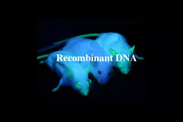
Transgenic Animals • Transgenic animals are produced by transferring cDNA into the pronucleus of a fertilized ovum. The resultant transgenic animal has the new gene (transgene) in all of its cells including its reproductive tissues. Transgenic animals are now widely used as experimental models in which to study human diseases. • A variation of this technique produces a knockout animal, in which a normal gene has been functionally eliminated. This may be done by site-specific mutagenesis. • Transgenic animals have a new gene (transgene) introduced into their germline. • All cells of a transgenic animal contain the transgene. • Transgene is inherited by offspring. • Knockout animals have a normal gene intentionally inactivated/destroyed. • Transgenic and knockout animals are used as models of human disease.
Gene Therapy • When using gene therapy to correct genetic deficiencies in humans, the cloned normal gene is targeted only to the tissues giving rise to the major symptoms. • For instance about 50% of the cases of severe combined immunodeficiency (SCID) are caused by mutations in the gene for a subunit common to several interleukin receptors. • The approach to gene therapy has been to introduce a normal cloned gene into the patient's bone marrow cells that will subsequently divide and differentiate to produce T and B lymphocytes expressing the gene. • These patients subsequently have improved immune function. Because the cloned normal gene has not been introduced into reproductive tissues any children of the patient would inherit the defective allele.
Glybera is a gene therapy that is designed to restore the LPL enzyme activity required to enable the processing, or clearance, of fat-carrying chylomicron particles formed in the intestine after a fat-containing meal. • The product consists of an engineered copy of the human LPL gene packaged with a tissue-specific promoter in a non-replicating AAV1 vector, which has a particular affinity for muscle cells. • The company produces Glybera using its insect cell-based manufacturing process. • Clinicians administer Glybera in a one-time series of up to 60 intramuscular injections in the legs.
Genome Editing Technologies Overview • The use of nucleases to make site-specific double-stranded breaks (DSBs) in the genome. Applications • To disrupt protein-coding genes and non-coding elements. Genetic variations can be created in cell lines and model organisms. • Can be injected directly into one-cell stage embryos to create gene knockout or knock‑in animals.
Facilitates the generation of disease models in various animals and in human pluripotent stem cells. • To enable the development of genetically improved crops and livestock. • Ex vivo and in vivo delivery of ZFNs have been used to edit genomes for the treatment of HIV infection in humans and haemophilia B in mice, respectively. • Gene correction and addition in patient-derived pluripotent stem cells or somatic cells using programmable nucleases can also provide novel therapeutic opportunities for patients with diverse genetic and acquired diseases.
ZFNs • Each “finger” recognizes about three or four base pairs of DNA • Three to six individual fingers can be linked to enable construction of arrays that recognize longer sequences of 9–18 base pairs (bp). • The nuclease domain of the zinc-finger is derived from the C-terminus of the FokI restriction endonuclease.
A schematic representation of a zinc-finger nuclease (ZFN) pair is shown. Each ZFN is composed of a zinc-finger protein (ZFP) at the amino terminus and the FokI nuclease domain at the carboxyl terminus.
TALENS (Transcription Activator Like Effectors): • TALENs are similar in architecture to ZFNs except that they use a different DNA-binding domain. • They consist of arrays of single protein modules that each recognize a single DNA base pair and that are derived from transcription activator like effectors (TALEs), factors encoded by plant pathogenic bacteria. • The nuclease domain used in TALENs is also from FokI
A schematic representation of a transcription activator-like effector nuclease (TALEN) pair is shown. Each TALEN is composed of transcription activator-like effectors (TALEs) at the amino terminus and the FokI nuclease domain.
CRISPR/CRISPR-associated (Cas) protein 9. CRISPR/Cas9 • Uses an RNA-guided system to perform genome editing. • gRNAs recognize 20-bp target sites • .
Nuclease-induced double-strand breaks (DSBs) can lead to sequence insertion, nucleotide correction or change (red box) through homology-directed repair (HDR) in the presence of a donor DNA or a single-strand oligodeoxynucleotide (ssODN), both of which contain homology arms. DSBs can also be repaired through error-prone non-homologous end-joining (NHEJ), which does not require donor DNA or ssODN and consequently often leads to small insertions and deletions (indels). Typical indel sequences and the number of inserted (+3 and +1) or deleted (–2, –4 and –10) bases are shown.