Download
1 / 53
530 likes | 537 Views
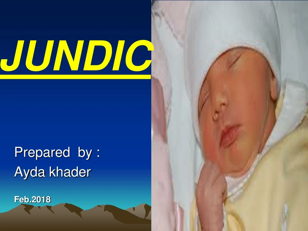
JUNDIC. E. Prepared by : Ayda khader Feb.2018. Objectives:. examine the physiological basis of neonatal jaundice Define the causes and consequences of pathological jaundice discuss the management of jaundice emphasize the role of the midwife in phototherapy

E N D
JUNDIC E Prepared by : Aydakhader Feb.2018
Objectives: • examine the physiological basis of neonatal jaundice • Define the causes and consequences of pathological jaundice • discuss the management of jaundice • emphasize the role of the midwife in phototherapy • emphasize the role of the midwife in preventing Rhesus isoimmunization
Jaundice • one of the most common conditions needing medical attention in newborn babies. • Is yellow coloration of the skin and the sclera caused by a raised level of bilirubin in the circulation (hyperbilirubinaemia). • Approximately 60% of term and 80% of preterm babies develop jaundice in the first week after birth, and about 10% of exclusively breastfed babies are still jaundiced at one month of age. • .
In most babies early jaundice is harmless • a few babies will develop very high levels of bilirubin, which can be harmful if not treated • Hyperbilirubinemia is exhibited as jaundice
Hyperbilirubinemia • In term neonates it appears when serum bilirubin concentrations reach (5–7 mg/dL), 85–120 μmol/L resulting from unconjugated bilirubin being deposited in the skin and mucous membranes .
Two main forms of bilirubin • Unconjugated bilirubin is fat soluble and cannot be excreted easily either in bile or urine. Neonatal jaundice can result from increased levels of this fat soluble bilirubin that cannot be excreted and is instead deposited in fatty tissue. • Conjugated bilirubin has been made water soluble in the liver and can be excreted in faeces and urine.Neonatal jaundice can also result from increased levels of this water soluble bilirubin if excretion is prevented by an obstruction.
Conjugation of bilirubin • Conjugation changes the end-products of red cell breakdown so they can be excreted in faeces or urine. • Understanding this process can increase evidence-based midwifery by increasing knowledge of the importance of such things as early breastfeeding, or early referral for treatment of pathological jaundice. • Ageing, immature or malformed red cells are removed from the circulation and broken down in the reticuloendothelial system (liver, spleen and macrophages). • .
Haemoglobin from these cells is broken down to the by-products of haem, globin and iron • Haem is converted to biliverdin and then to unconjugated bilirubin • Globin is broken down into amino acids, which are used by the body to make proteins • Iron is stored in the body or used for new red cells.
Three stages are involved in the process of bilirubin conjugation: 1-Transport of bilirubin • Unconjugated or fat soluble bilirubin is transported to the liver bound to albumin. If not attached to albumin, this unbound or ‘free’ bilirubin can be deposited in extravascular fatty and nerve tissues (skin and brain). • Skin deposits of unconjugated or fat soluble bilirubin cause jaundice, while brain deposits can cause bilirubin toxicity or kernicterus
2- Conjugation • Once in the liver, unconjugated bilirubin is detached from albumin, combined with glucose and glucuronic acid and conjugation occurs in the presence of oxygen and the enzyme Uridine diphospho glucuronyl transferase (UDP-GT). • The conjugated bilirubin is now water soluble and available for excretion.
3- Excretion • Conjugated bilirubin is excreted via the biliary system into the small intestine where normal bacteria change the conjugated bilirubin into urobilinogen. • This is then oxidized into orange-colouredurobilin. • Most is excreted in the faeces, with a small amount excreted in urin
Jaundice Jaundice is caused by bilirubin deposits in the skin. In term neonates it appears when serum bilirubin concentrations reach 85–120 μmol/L (5–7 mg/dL), with a head to toe progression as levels increase.
Physiological jaundice • Neonatal physiological jaundice occurs when unconjugated (fat soluble) bilirubin is deposited in the skin instead of being taken to the liver for processing into conjugated (water soluble) bilirubin that can be excreted in faeces or urine. • It is affecting up to 50% of term and 80% of premature babies who have a progressive rise in unconjugated bilirubin levels and jaundice on day 3. • Physiological jaundice never appears before 24 hrs of life • usually fades by 1 week of age and bilirubin levels never exceed 200–215 μmol/L (12–13 mg/dL).
Factors that contribute its: • relative Polycythemia • a shortened red blood cell lifespan, • immature hepatic uptake and conjugation process • increased enterohepatic circulation
Early physiological jaundice (within first 5 days after birth)Possible causes include: • physiological jaundice • haemolysis (Rhesus isoimmunization, ABO incompatibility, other blood group antigen problems) • infection • bruising • polycythaemia • dehydration (unlikely in the first 48 hours but must be considered in babies presenting between 2–7 days after birth, particularly those who are breast fed).
Assessment and diagnosis of physiological jaundice: The initial assessment of a baby should include identifying risk factors for jaundice. These include any disease or disorder that increases bilirubin production, or alters the transport or excretion of bilirubin.
For example: • birth trauma or evident bruising (increased production of unconjugated bilirubin) • family history of significant haemolytic disease or jaundiced siblings • maternal antibodies at booking • evidence of infection • prematurity • timing of jaundice, for example, within the first 24 hours (suggesting haemolysis).
Pathologic jaundice Pathological jaundice in newborns usually appears within 24 hrs of birth, and is characterized by a rapid rise in serum bilirubin. • jaundice within the first 24 hrs of life • total serum bilirubin > 200 μmol/L (12 mg/dL) • persistence of clinical jaundice for 7–10 days in term or 2 weeks in pre-term babies.
Risk Factors: • Conditions that alter the production, transport, uptake, metabolism, excretion, or reabsorption of bilirubin can cause pathologic jaundice newborn • Polycythemia, blood incompatibilities, and systemic acidosis. • These altered conditions can lead to high levels of unconjugated bilirubin, possibly reaching toxic levels • and resulting in a severe condition called Kernicterus.
Kernicterus (bilirubin encephalopathy): • Is a preventable neurologic disorder characterized by encephalopathy, motor abnormalities, hearing and vision loss, and death. • Neurotoxicity develops because unconjugated bilirubin has a high affinity for brain tissue, and bilirubin not bound to albumin is free to cross the blood-brain barrier and damage cells of the CNS.
Diagnostic test : • serum bilirubin to determine levels and if bilirubin is unconjugated or conjugated • Total serum protein to detect reduced binding capacity of albumin • Direct Coomb's test to detect presence of maternal antibodies on fetal RBCs • indirect Coomb's test to detect the presence of maternal antibodies in serum • reticulocyte count – elevated by haemolysis as new RBCs are produced 6) ABO blood group and Rh type for possible incompatibility 7) haemoglobin / haematocrit estimation to assess anaemia
Treatment strategy These include phototherapy, Immunoglobulin , exchange transfusion.
Phototherapy • reduces levels of unconjugated bilirubin in the blood and decreases the likelihood of neurotoxicity or kernicterus. • The skin surface is exposed to high intensity light, which converts fat-soluble unconjugated bilirubin into water-soluble bilirubin that can be excreted in bile and urine. Commercially available phototherapy systems include those delivering light via fluorescent bulbs, halogen quartz lamps, light-emitting diodes and fibreoptic mattresses
Indications for phototherapy based on serum bilirubin levels and the individual condition of each baby, particularly when jaundice occurs within the first 12–24 hrs: • for pre-term infants <1500 g – between 85 and 140 μmol/L (5 and 8 mg/dL) • for pre-term infants >1500 g, sick infants and those with haemolysis – between 140 and 165 μmol/L (8 and 10 mg/dL) • for healthy term infants jaundiced after 48 hrs – between 280 and 365 μmol/L (17 and 22 mg/dL).
Consideration of the above individual factors, and serum bilirubin levels <215 μmol/L (13 mg/dL) are usual before stopping phototherapy. • Although bilirubin levels can rise following phototherapy, healthy term babies do not require testing to identify this rebound effect. Rebound to clinically significant levels is more likely with prematurity, a positive direct Coombs test, and in those treated before 72 hrs
Stopping phototherapy • The SBR should be measured at least every 6–12 hours whilst phototherapy continues. It should be monitored more frequently when the rate of rise is rapid. • Phototherapy may be safely discontinued when the bilirubin is 50 µmol/l below the threshold. • Repeat SBR measurement is necessary 12–18 hours after ceasing phototherapy to check for rebound hyperbilirubinaemia.
Complication of phototherapy • hyperthermia, increased fluid loss and dehydration • damage to the retina from the high intensity light • lethargy or irritability, decreased eagerness to feed, loose stools • skin rashes and skin burns • alterations in a baby's state and neurobehavioral organization • isolation and lack of usual sensory experiences, including visual deprivation
7) decrease in calcium levels leading to hypocalcaemia 8) low platelet counts and increased red cell osmotic fragility 9) bronze baby syndrome and DNA damage.
Midwifery care and phototherapy Midwives and family are usually responsible for infant care either in hospital or at home • The infant is maintained in a warm thermoneutral environment and observed for hypo- or hyperthermia. • Eye shields or patches must cover the eyes without occluding the nose • Skin is cleaned with warm water and observed for rashes, dryness and excoriation. • Fluid intake and output are monitored and demand feeding is continued.
Babies receiving phototherapy should be , a minimum of 40 cm from the light • phototherapy equipment should be routinely checked for safety • temperature should be measured and recorded at least 4-hourly • Application of topical creams or lotions should be avoided as there is a risk of burns and blistering • The baby should be assessed regularly for signs of dehydration
can be removed/switched off during cares and feeds (for up to 30 minutes in every 3 hour period is acceptable while on single phototherapy. • if the baby is requiring multiple phototherapy this should not be interrupted. • Monitor for Neurobehavioural status. This includes sleep and wake states, feeding behaviours, responsiveness, response to stress and interaction with parents and other carers. • Parent support, parents will be caring for their infant and need adequate information
Exchange transfusion Excess bilirubin is removed from the baby during a blood exchange transfusion. In recent years, cord blood screening and advances in phototherapy have reduced exchange transfusion for infants with many haemolytic and enzyme deficiency diseases. Except with very premature babies and Rh incompatibility, exchange transfusion may only be used when phototherapy has failed, or there is a risk of kernicterus.
It is an ideal dilution of serum bilirubin and antibodies. • A catheter is introduced into the umbilical vein after cutting the cord. • It involves transfusing a large volume of blood to the baby (double the baby's blood volume or 160 ml/kg) • Exchange is carried out over 45-60 min period by alternating aspiration of 20 ml of newborn's blood and infusions of 20 ml of the donor blood
Complications: 1. Embolism, thrombosis, infarction. 2. Arrhythmias, heart failure. 3. Electrolyte disturbances. 4. Thrombocytopenia. 5. Infections. 6. Hypo and hyperthermia.
Midwifery responsibilities: • Keep the newborn NPO for 2-4 hours before exchange to prevent aspiration. • Check donor blood for compatibility. • Keep resuscitation equipment at beside: oxygen, ambo bag, endotracheal tubes, and laryngoscope. • Assist physician with exchange transfusion procedure. • Track amount of blood withdrawn and transfusion to maintain balanced blood volume
Maintain body temperature to avoid hypothermia and cold stress. • Monitor vital signs and observe for rash. • after transfusion, continue to monitor vital signs and check umbilical cord for bleeding or signs of infection.
Haemolytic jaundice • increased haemoglobin destruction in the fetus or newborn has several cause: • Rhesus (RhD) isoimmunization • ABO incompatibility. This increased haemolysis increases bilirubin levels, and causes pathological jaundice. .
Rhesus (RhD) isoimmunization • can occur if blood cells from a Rhesus-positive baby enter a Rhesus-negative mother's bloodstream. • Her blood treats the D antigen on positive blood cells as a foreign substance and produces antibodies • the midwife has critical role in the injection of anti-D immunoglobulin (anti-D Ig). • Without this anti-D prophylaxis, RhDisoimmunization can cause severe haemolytic disease of the newborn (HDN) with significant mortality and morbidity
ABO incompatibility • ABO isoimmunization usually occurs when the mother is blood group O and the baby is group A, or less often group B. • Type O women are 5.5 times more likely to have sensitization than type A or B as the latter have a protein or antigen not present in type O blood. • Individuals with type O blood develop antibodies throughout life from exposure to antigens in food, Gram-negative bacteria or blood transfusion • the first pregnancy may already have high serum anti-A and anti-B antibody titres. • .
Some women produce IgG antibodies that can cross the placenta and attach to fetal red cells and destroy them • ABO incompatibility is also thought to protect the fetus from Rh incompatibility as the mother's anti-A and anti-B antibodies destroy any fetal cells that leak into the maternal circulation
Although first and subsequent babies are at risk, destruction is usually much less severe than with Rh incompatibility. • In most cases haemolysis is fairly mild but in subsequent pregnancies can become more severe. • ABO erythroblastosis can, rarely, cause severe fetal anaemia and hydrops
if babies require phototherapy it is usually commenced at a lower serum bilirubin level (140–165 µmol/l or 8–10 mg/dl). • In rare cases, babies with high SBR levels require exchange transfusion. • IVIG administration to newborns with significant hyperbilirubinaemia due to ABO haemolytic disease (with a positive direct Coombs' test) has reduced the need for exchange transfusion
Late neonatal jaundice • This is generally defined as abilirubin concentration that remains raised beyond 14 days of age. • There are a number of causes and investigation is important because, whilst uncommon, some of the causes are conditions that have significant long-term implications if not treated and treatments are available which are effective if used early enough.
Causes of late neonatal jaundice • Any disease or disorder that increases bilirubin production or alters transport or metabolism of bilirubin is superimposed upon normal physiological jaundice. • It is best to divide the causes into those conditions that cause a raised : • unconjugatedbilirubin (fat soluble) • conjugated bilirubin (water soluble).
Late neonatal (>14 days) unconjugated hyperbilirubinaemia: • Increased red cell destruction or haemolysis causes raised SBR levels and blood type/group incompatibility, including Rhesus (Rh-D) and ABO incompatibility • Other factors include sepsis, particularly urinary tract infection, hypothyroidism and galactosaemia. Non-immune haemolysis features spherocytosis (fragile red cell membranes) and enzyme deficiencies. Glucose-6-phosphate dehydrogenase (G6PD)
G6PD is an enzyme that maintains the integrity of the cell membrane of RBCs and deficiency results in increased haemolysis. • G6PD deficiency is an X-linked genetic disorder carried by females that can affect male babies of African, Asian and Mediterranean descent.


![{90K} [Bilal] : "A New Breed of Hero" (2018) s-t-r-e-a-m-i-n-g english](https://cdn4.slideserve.com/7769521/bilal-a-new-breed-of-hero-2018-full-movie-online-dt.jpg)