Download
1 / 115
1.16k likes | 1.39k Views
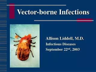
Tick-borne Infections. Pola de la Torre, M.D. Assistant Professor of Medicine Division of Infectious Diseases. Ticks. Blood-sucking arthropods (eight legged) of the class Arachnida. Three families: -Ixodidae (hard ticks) -Argasidae (soft ticks)

E N D
Tick-borne Infections Pola de la Torre, M.D. Assistant Professor of Medicine Division of Infectious Diseases
Ticks • Blood-sucking arthropods (eight legged) of the class Arachnida. • Three families: -Ixodidae (hard ticks) -Argasidae (soft ticks) -Nuttalliellidae (characteristics of both)
Ixodes • Three-stage life cycle -Larva -Nymph -Adult • Require a blood meal at each stage. • Peak questing periods -Larvae: August and September. -Nymphs: May through July. -Adults: Spring and Fall.
Ticks • The host’s epidermis is penetrated by the tick’s chelicerae. • The resulting defect guides the tick’s hypostome into place to begin withdrawal of blood. • Hard ticks can secrete a liquid cement from salivary glands so that it can remain in place for the 7-14 days required for a blood meal. • After several hours of attachment disease transmission can take place.
Removing Attached Ticks • Use forceps and detach the intact tick without leaving the mouth parts in the skin. • Monitor for signs and symptoms of tick-borne diseases for up to 30 days.
The Pathogen • Causative agent -Rickettsia rickettsii -Nonmotile pleomorphic weakly gram-negative cocobacillus - Intracellular obligate that resides more in the cytosol (less so in the nucleus) of host cells. -Can be seen with Gimenez or acridine orange stains.
Epidemiology • In 1908 the role of a tick bite in transmitting RMSF was described. • The seasonal distribution of disease parallels tick activity. • The tick is both the vector and the main reservoir. • Most cases are diagnosed in late spring and summer.
Epidemiology • Dermacentor variabilis -Two thirds of US and the Far West • Dermacentor andersoni -Western states • Rhipicephalus sanguineus -Mexico • Amblyomma cajennense -South America
Pathogenesis • Rickettsiae enter through the skin and spread via the lymphatics and small blood vessels to the systemic and pulmonary circulation. • They attach to (Omph A, Omph B, rickettsial phospholipase) vascular endothelium. • After induced phagocytosis, rickettsiae escape from the phagosome into the cytosol.
Pathogenesis • Rickettsiae proliferate intracellularly by binary fission and are released to infected adjacent cells (the maculopapular rash). • The effect of this endothelial cell injury is increased vascular permeability which results in edema, hypovolemia, hypotension, and hypoalbuminemia. • Hyponatremia results from secretion of ADH in response to the hypovolemia.
Pathogenesis • Vascular injury and the subsequent host response correspond to the distribution of rickettsiae. -Interstitial pneumonia, interstitial myocarditis, perivascular glial nodules of the CNS and similar vascular lesions in the rash, GI tract, pancreas, liver, skeletal muscles, and kidneys.
Pathogenesis • Significant hemorrhage is rare. • Platelets are consumed in the localized lesions and thrombocytopenia can be seen in 32-52% of patients. • A procoagulant state occurs but true DIC is rare and occlusive vascular thrombosis is not the basic pathophysiologic event.
Clinical Manifestations • Incubation period: 2-14 days (median 7d). • Usually begins with fever, myalgia, and HA. • Temp >102 F in 63% during the first 3 days and in 90% later. • Other S/S frequent early before the onset of rash: nausea, vomiting, abdominal pain, diarrhea, abdominal tenderness.
Clinical Manifestations • The major diagnostic sign-The Rash: -Small fraction on the first day. -49% during the first 3 days. -Usually appears 3-5 days after the onset of fever and occurs in 84-91% overall. • Rash typically begins around the wrists and ankles but may start on the trunk or be diffuse on the onset.
Clinical Manifestations • Involvement of the palms and soles often appears late and although thought to be characteristic occurs in only 36-82% with rash. • Skin necrosis or gangrene in 4%. • Finding a eschar at the site of the bite rare.
Clinical Manifestations • HA quite severe. • Focal neurologic deficits, transient deafness, meningismus, and photophobia may suggest meningitis or meningoencephalitis. • CSF: 1/3 have increased leukocytes, elevated protein in 1/3, glucose low in only 8%. • Generally, neurologic involement portends a bad prognosis.
Clinical Manifestations • Retinal vein engorgement, arterial occlusion, flame hemorrhage, and papilledema without increased CSF pressure. • Prerenal azotemia. • ATN. • Pulmonary edema.
Clinical Manifestations • Classic RMSF -Without appropriate therapy death occurs 8-15 days after onset of symptoms. -Labs nonspecific: WBC generally WNL but with increased immature myeloid cells, anemia in 5-30%, thrombocytopenia, coagulopathy is infrequent. -Hyponatremia in 50%. -Increased LDH, CK, other enzymes related to diffuse tissue injury.
Clinical Manifestations • Fulminant RMSF -Death within the first 5 days -Diagnosis difficult Antibodies haven’t developed Characteristic lesions appear different -Black males with G6PD deficiency, older age, possibly alcoholism.
Diagnosis • Before the onset of rash the diagnosis is clinical and epidemiologic. • Differential Dx: typhoid fever, measles, rubella, respiratory tract infection, gastroenteritis, acute abdomen, enteroviral infection, meningococcemia, disseminated GC, secondary syphilis, leptospirosis, immune complex vasculitis, ITP, TTP, EBV, drug reaction, ehrlichioses, other rickettsial diseases.
Diagnosis • Serology (retrospective) -A fourfold increase in titer between acute and convalescent stages is diagnostic. -A titer of ≥64 detectable between 7 and 10 days after the onset of illness.
Treatment • Doxycycline 100 mg q12 hours • Chloramphenicol 50-75 mg/kg/day • Usually 7 days and continued for 2 days after the patient has become afebrile.
The Pathogen • Tick-borne spirochete Borrelia burgdorferi • Lyme disease or borreliosis was recognized in 1976 following a cluster of affected children in Lyme Connecticut were thought to have juvenile rheumatoid arthritis.
The Pathogen • After injection of B.burgdorferi by the tick and an incubation period of 3-32 days, the organism first multiplies at the site of the bite in the skin. • Within days to weeks the organism disseminates through the body.
Clinical Manifestations • Early Infection: Stage 1(Localized) -About 70-80% develop EM (expands slowly at 1 cm/day) at the site of the bite. -Most patients do not remember the bite because the small size of the nymphal I. scapularis.