Download
1 / 27
270 likes | 569 Views
Evaluation of Thyroid Nodules. Michael L. Tuggy, MD Swedish Family Medicine, Seattle, WA. Case 1. 42 y.o. male with no active medical problems. During your routine physical, note a thyroid nodule. Told by ENT last year not to worry about it. PE: 1 x 2cm R lower pole nodule.

E N D
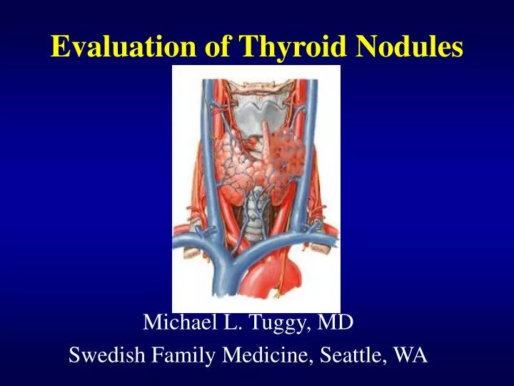
Evaluation of Thyroid Nodules Michael L. Tuggy, MD Swedish Family Medicine, Seattle, WA
Case 1 • 42 y.o. male with no active medical problems. During your routine physical, note a thyroid nodule. Told by ENT last year not to worry about it. • PE: 1 x 2cm R lower pole nodule. What information do you want from the patient?
Age as a Risk Factor • Age • young patients (<20 years of age) • thyroid nodules are much more likely to be malignant (40-50%). • elderly (>60 years of age) -higher risk, especially of more aggressive thyroid tumors.
Gender and Thyroid Nodules • Gender • male -higher risk if nodule present • females • have many more nodules • less likely to be malignant. • still have majority of thyroid cancers
Other major risks • Radiation to head and neck. • 40% risk of thyroid cancer usually 25 years later. • Exposed populations- Polynesian studies • Family History of MEN II, Gardner’s Syndrome, Cowden’s disease.
Historical Red Flags • Recent growth • Soft tissue swelling • Vocal changes • Dysphagia • Signs of thyroid dysfunction
Case 2 • 26 y.o. Eritrean female with a 2-3 year history of goiter. No symptoms but noted enlargement on right for 1 year. • P.E.: 3x4 cm Right sided thyroid mass, firm, adherent to soft tissue. What physical findings are worrisome? How can you best clarify the nature of the nodule?
Physical Exam of the Thyroid • Use both hands simultaneously to evaluate for symmetry • Patient upright - screening exam • Patient supine with neck in extension- detailed exam. Swallowing assists in elevating gland. • Evaluation of other neck structures. • Voice changes (recurrent laryngeal nerve).
Thyroid Scans • Purpose • Determine function of the gland and/or a nodule within the gland • Hot nodules - usually independently functioning nodules • Rarely, rarely malignant • Cold nodules - either adenoma or maligancy • 15% chance of malignancy in adults.
Thyroid Ultrasound • Can identify presence of nodules. • May be able to characterize follicular vs. solid. • Not able to rule our malignant nodule • Aid in biopsy. Thyroid
Case 3 • 30 y.o. WF with enlarging cold benign thyroid adenoma (diagnosis from previous FNA biopsy). • PE: 4 x 5 cm mass on Right What do you do now?
Fine-Needle Aspiration • Best tool for determining pathology other than surgical excision. • Can be as high as 80 % sensitive and 95% specific. • Operator dependent in obtaining adequate amount of tissue. 25 gauge needle is optimal. • Should not be relied on if negative in patient with previous neck irradiation. • Multifocal tumors common.
Interpreting the Biopsy Report • What you get: • benign • indeterminate • suspicious • inadequate specimen • What it means: • benign - 90-95% likelihood it is benign • indeterminate- who knows? • suspicious- it’s malignant. • inadequate specimen - do it again (and again)
Thyroid Malignancies- Papillary • Most common • 30% have node metastasis at diagnosis • Radiation related • Histologically, psammoma bodies distinguish from benign adenoma.
Thyroid Malignancies-Follicular • 20 % of malignancies • Distinguished from normal follicular adenomas by invasion of capsule or blood vessels. • May be difficult to determine on FNA
Thyroid Malignancies- Medullary • 5-10% of cases • arise from the C cells which produce calcitonin • diagnosis based on elevated thyrocalcitonin levels and thyroid nodule (cold)
Thyroid Malignancies- Anaplastic • < 10% • Highly aggressive with local extension at time of diagnosis. • No suitable therapy • Prognosis < 1 yr from diagnosis
Treatment • For all malignancies, excision of the the lobe (or if post-radiation the entire gland). • XRT- very specific and well tolerated- I131 therapy. • Anaplastic tumors - palliative radiation and XRT.
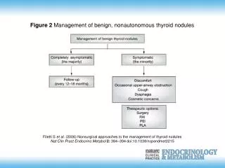
What about those benign nodules? • No specific treatment is needed. • Thyroid suppression may shrink size of adenomas • Not proven to be effective or necessary • May hide malignancies - ? Periodic biopsies or scans.
Case 4 - This weeks puzzler! • 40 y.o. WF s/p I131 ablation for Grave’s Dz. 6 years ago. • Persistant R thyroid nodule 2 x 1.5 cm in size. What is the likely diagnosis?
Outcomes • Case 1. - Papillary cancer - 3 (+) nodes • no metastasis at 1 year. • Case 2. - Follicular cancer - 5 (+) nodes • no metastasis at 1.5 years • Case 3. - Large adenoma with incidental 1 cm papillary carcinoma superior to nodule. • No recurrence at 5 years. • Case 4. - Non-functional adenoma
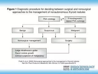
Modified from: Castro, MR, Gharib, H. Endocr Pract 2003; 9:128.
Summary:Solitary Nodule Evaluation • TSH – if low – scan – if hot nodule, then observe. • Normal TSH - Do I scan first or FNA first?- • high risk - scan and FNA • Is the nodule cold or hot? • Cold - FNA biopsy • low risk - FNA • if indeterminate- scan and re-FNA or excisional biopsy. • Anti-perioxidase Antibody – helpful if low- TSH to diagnose thyroiditis.

![[PDF Read❤️ ONLINE] Thyroid Cancer and Thyroid Nodules In 30 Minutes: A guide to s](https://cdn7.slideserve.com/12662172/slide1-dt.jpg)