Download
1 / 30
300 likes | 505 Views
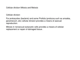
Cellular Division. Binary Fission. DNA replicated. Membrane added. Cell Division. Functions in Reproduction, Growth, and Repair Unicellular Organisms Reproduction of Entire Organisms Multicellular Organisms Growth and Development from the Fertilized Egg

E N D
Binary Fission DNA replicated Membrane added
Cell Division • Functions in Reproduction, Growth, and Repair • Unicellular Organisms • Reproduction of Entire Organisms • Multicellular Organisms • Growth and Development from the Fertilized Egg • Replacement of Damaged or Dead Cells • Distributes Identical Sets of Chromosomes • Precisely Replicates its DNA • Results in 2 Daughter Cells
Binary Fission in Bacterium:Asexual Reproduction by Mitosis Nuclearmaterial Division plane Cell wall Cytoplasm
Binary Fission in Paramecium:Asexual Reproduction by Mitosis New individuals
Budding Yeast:Asexual Reproduction by Mitosis • Nucleus divides by mitosis. • Bud forms on cell. • Nucleus moves into bud. • Bud separates.
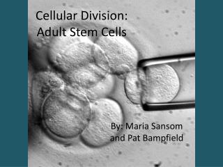
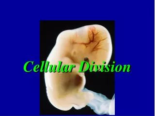
Growth and Development:Functions of Mitosis Organs Fertilized egg (zygote) Multicell stage Mitotic cell division & differ-entiation Mitotic cell division Tissues
The EukaryoticCell Cycle G1: Growth cytokinesis telophase S: Synthesis of DNA; chromosomes duplicated anaphase celldivision Mitosis metaphase prophase interphase G2: Growth G0: nondividing
The Cell Cycle • Alternates between mitotic (M) phase and interphase in eukaryotes. • Interphase • ~90% of cycle • chromosome replication • cell growth and metabolism • three periods • G1 phase – first growth phase • S phase – synthesis of duplicated DNA • G2 phase – second growth phase
Interphase (nondividing) Interphase : The chromosomes (blue) are in the thin, extended state and appear as a mass in the center of the cell. The microtubules (red) extend outward from the nucleus to all parts of the cell.
Phases of Mitosis (nuclear division): Prophase Interphase : The chromosomes (blue) are in the thin, extended state and appear as a mass in the center of the cell. The microtubules (red) extend outward from the nucleus to all parts of the cell. Late prophase: Chromosomes have condensed and attached to microtubules. Microtubules have reorganized to form the spindle. Chromatid develops a kinetechore.
Chromosome Condensation achromatid centromere single-stranded chromosome double helixuncondensed double-stranded chromosome double helicesstill uncondensed DNA replication Chromosome condensation cell 1 double-stranded chromosome 2 double helicesnow condensed basepairs closer look still closer look even closer look
Human Karyotype, Male • These are chromosomes from mitosis • Stained to show regions • Numbered by length • Occur in pairs
Phases of Mitosis (nuclear division):Metaphase Interphase : The chromosomes (blue) are in the thin, extended state and appear as a mass in the center of the cell. The microtubules (red) extend outward from the nucleus to all parts of the cell. Late prophase: Chromosomes have condensed and attached to microtubules. Microtubules have reorganized to form the spindle. Chromatid develops a kinetechore. Metaphase: The chromosomes have moved along the spindle microtubules to the equator of the cell. Centromeres align on the equator. Entire structure of microtubules is called the spindle.
Separation of Sister Chromatids • In metaphase, sister chromatids are held together at centromere • At end of metaphase, centromere releases sister chromatids • In anaphase, they move to opposite poles
Phases of Mitosis:Anaphase & Telophase Anaphase: Sister chromatids have separated into separate chromosomes. Kineteochore microtubules shorten at kinetochore. Poles move farther apart. Telophase: Nonkinetochore microtubules elongate cell. The chromosomes have gathered into two clusters, one at the site of each future nucleus. Next interphase: Chromosomes are relaxing again into their extended state. Spindle fibers are disappearing, and the microtubules of the 2 daughter cells rearrange into the interphase pattern.
Mitosis:Prophase - Metaphase Nucleolus disappears; Nuclear envelope breaks down Duplicated chromosomes remain elongated Chromosomes condense and shorten LateInterphase EarlyProphase LateProphase Metaphase Centrioles have also been duplicated Centrioles begin to move apart; Spindle forms Microtubules attach to kinetochores Kinetochores align at cell’s equator
MitosisAnaphase - Cytokinesis One set of chromosomes; Begin unwinding Free spindle fibers push poles apart Cytoplasm divided along equator NextInterphase Anaphase Telophase Cytokinesis Each daughter gets 1 nucleus & half of cytoplasm Spindle disappears; Nucleolus reappears Chromatids become independent chromosomes Nuclear envelope re-forms
Cytokinesis of a Ciliated Cell Daughter Cells Cleavage Furrow
Cytokinesis in Plants Vesicles fuse to form cell wall and membranes Complete separation of daughter cells
Meiosis I Homologous chromosomes exchange DNA & align on equator Homologous chromosomes move to opposite poles Homologous chromosomes pair and cross over Telophase I Prophase I Metaphase I Anaphase I
Meiosis II Similar to Mitosis FourHaploidCells Prophase II Metaphase II Anaphase II Telophase II
Crossing Over • Homologues pair up • Protein strands zip together • Recombination enzymes snip and rejoin DNA • Homologs separate with new gene combinations
Meiosis vs. Mitosis:Comparison of Spindles Meiosis: Duplicated chromosomes with one kinetochore; Paired homologues go to opposite poles. Mitosis: Duplicated chromosomes with two kinetochores; Unpaired homologs split between sister chromatids, which go to opposite poles.