Download
1 / 59
760 likes | 1.54k Views
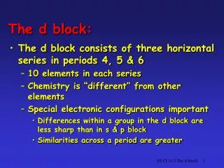
Pediatric Ultrasound guided blocks “ block under vision “. Dr. Amr Abdulfatah Sayed (M.D.) Associate prof. of Anesthesia, Chronic Pain Management Ain Shams Univ. , Cairo , EGYPT Oct. 2012. Imagine . NO clicks No pops No paresthesia No trans-arterial . Under vision in real time

E N D
Pediatric Ultrasound guided blocks“block under vision “ Dr. Amr Abdulfatah Sayed (M.D.) Associate prof. of Anesthesia, Chronic Pain Management Ain Shams Univ. , Cairo , EGYPT Oct. 2012
Imagine • NO clicks • No pops • No paresthesia • No trans-arterial • Under vision in real time • How L.A. behaves • How catheter lodge • Reinjection with inconsistent block
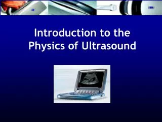
Basic Principles • Mechanical sound energy. • Sinusoidal. • Pulse longitudinalwavealternating compression (high pressure) & rarefaction (low pressure) • P = pressure • T = wave length • F= frequency • distance one peak to other peak is a wavelength one peak to other peak is a wavelength
GENERATION OF AN ULTRASOUND WAVES • electric field is applied to a piezoelectric crystals. • mechanical distortion of the crystals • sound waves (i.e. mechanical energy)
Ultrasound & Tissue Interaction • U/S beam travels through different tissues • Subjected to attenuation(Energy Loss). 1) absorption.2) reflection.3) scattering. • Factors affecting attenuation : • Frequency ( high highatten ) ( low lowatten) • Travel distance • Tissue nature
Fact The Ultrasound Transducer Source of Energy and Image Energy progressively degraded (attenuate)as it enters deeper tissues lateral & axial resolution improved with higher frequency transducers,decrease with increasing tissue depth
Facts • Structures typically seen as hyperechoic or echogenic include bone, tendons, pleura, and nerves below the clavicles. • In contrast, blood, fluids, and nerves above the clavicles are hypoechoic.
Facts • Generally speaking, a high frequency wave is subjected to high attenuation thus limiting tissue penetration • low frequency wave is associated with low tissue attenuation and deep tissue penetration.
GAIN & TIME GAIN COMPENSATION (TGC) • receiver amplification is called the Gain. • Gain increases overall brightness of the entire image, including the background noise. • (TGC) selectively amplify the weak returning (attenuated) signals from deeper structures. TGC Gain
TISSUE ECHOGENICITY • Tissue impedance • resistance of a tissue to US passage • Strong wave reflection = hyperechoic (white) • Weak reflection= hypoechoic (greyish) • No reflection = anechoic. (black)
TISSUE ECHOGENICITY • Bone : High tissue impedance • Strong reflection • ++ hyperechoic lines with a hypoechoic shadow underneath
Neural Tissue Echogenicity fascicular or honeycomb appearance
Reflection is high for air • Air has extremely low acoustic impedance (0.0004) • acoustic coupling medium on the transducer surface to eliminate any air pockets • Otherwise ultrasound waves will be reflected limiting tissue penetration. large dropout artifact.
Acoustic Shadow Artifact • deep to hyperechoic bone outline is a beam attenuation • Ultrasound beams subjected to attenuation by bone, penetration is severely impeded.
Linear array probe • High frequency( > 6MHz) • Superficial structures • Depth max. 6 cm • High clarity • Curved probe • Low frequency (2-5 MHz) • Deep structures > 6cm • Less resolution Hokey stick 25mm
Probe Orientation • Transducer marker
Practical Recommendations • Needle probe orientation • Handling of probe resting hand on pt. body • Non dominant hand • More steep angle of needle = difficult visualization • IN PLANE (IP) • OUT OF PLANE (OOP)
The more perpendicular the needle is to ultrasoundbeam, the stronger (brighter and solid) the needle will appear on the monitor
Needle Selection • Large bore needles (e.g. 17 G) • Better visualization • easier to direct. • Preferred for deep blocks (e.g. infraclavicular block, sciatic ) when needle insertion is steep (> 45 degrees) • Smaller bore needles (e.g. 22 G) • easily visualized for more superficial blocks e.g., the axillary block, when the angle of needle insertion is shallow.
Movement of Probe (ART) • A ( Alignment ) • R ( Rotation) • T ( Tilting)
Proper Body Ergonomics • Proper handling the transducer and the needle, to view the screen, and to position the patient are essential for block success and to avoid operator fatigue and body injury.
Proper Body Ergonomics Improper body position Proper body position
Proper Body Ergonomics Proper operator and screen orientation Improper operator and screen orientation
Proper Body Ergonomics Improper hand and arm positions Proper hand and arm position
Local Anesthetic Injection • Aim to surround the neural structure • Doughnut sign. • Saline of D5% ( if PNS) Prior to L.A • Aspirate 1st
Femoral Nerve • doughnut” sign. F.A F.A Doughnut sign.
Glut. max Sc. N.
Ultrasound With & Against proponents critics • improve success rates • simplify the technical challenges • decrease performance times • reduce complications • lack of controlled trials • High expense • equivocal nature of images • Requires sustained training.
Pediatric USGRA Tips • Adults USGRA can be performed in children • Plan @ pre-op visit • Explain for partents ( legal guardian ) • Ped. USGRA performed under anesthetized • light sedation ( esp. > 8 yrs , diff. airway ,MH). • EMLA 1 hrs in advance • Small muscle bulk = high f probe (13-6 MHz, “hockey-stick,” 26-mm footprint)
Pediatric USGRA Tips • Insulated needle • Epidural 22G for catheter ( long term post op). • In-plane > out off plane • Calculate full dose ( volume 0.3–0.5 mL/kg ) • < 5 yrsBupi. 0.25 %, Ropi. 0.2%, Lido 1% • 5 yrsBupi. 0.375 %, Ropi. 0.5% +epi. (Allison Ross et al. , A & A July 2000 vol. 91 no. 1 16-26)
should we use nerve stimulation in addition to U/S Guidance?
Upper Limb Blocks • Interscalene • Supraclavicular • Infraclavicular • Axillary • Median N. • Ulnar N. • Radial N. 0.25 -0.5ml /kg 0.25% bupivacaine , 1% lidocaine , 0.25% ropivacaine
Interscalene • Most cranial approach • Not popular ( Phrenic N.) • Indication : shoulder , upper arm, lateral clavicle. • Scanning : • Medial to lateral survey • Trace Back Method • Injectate “Perfect Block”= 0.15-0.3ml /kg front, in the sheath& behind
Trace Back SCM Sc. m Sc. a medial ColourDoppler : to identify vertebral vs & branches of transverse cervical artery below C6
Supraclavicular (Spinal Of UL) • Clinical tips • The usual volume of L.A. 0.3-0.5ml/kg. • (Corner Pocket ) above the 1st rib, next to subclavian a. to anesthetize the lower trunk. • In plane approach is a must • All U.L • ???? Shoulder surgery • 2/3 L.A. @ Corner
Infraclavicular (Cords) Local anesthetic injected posterior to the axillary artery resulting in a U shape spread around the artery is associated with complete blockade of the arm, forearm and hand.
Axillary Approach (terminal branches of the brachial plexus ) • Below elbow surgeries AA MCN Inject : MCN +12 & 6 O’clock Inject : MCN + 10 , 2 , 6 O’clock In Plane
Femoral C superficial to the Ilio-Psoas Muscle (IPM) Base touching CFA, extending lateral to it.
Femoral Surgery on femur , knee