Download
1 / 67
700 likes | 889 Views
LYMPHATIC SYSTEM. Chapter 14. The lymphatic system is part of the circulatory system. Two key roles in the body: Carries fluid from the extracellular environment into the bloodstream, and thereby helps to maintain fluid balance.

E N D
LYMPHATIC SYSTEM Chapter 14
The lymphatic system is part of the circulatory system. • Two key roles in the body: • Carries fluid from the extracellular environment into the bloodstream, and thereby helps to maintain fluid balance. • It performs the primary role in defending the body against disease.
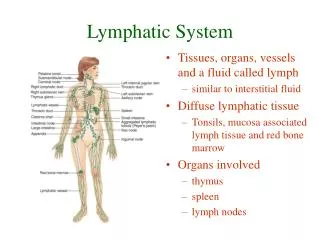
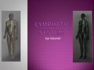
Specialized organs of Lymphatic System • Lymph nodes, the spleen, the thymus, the tonsils, and Peyer’s patches
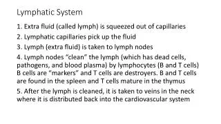
The Lymphatic Network • Interstitial fluid is formed when plasma from the bloodstream is pushed through capillary walls. • This watery fluid serves as a transport medium for gases, nutrients, and other molecules that travel between cells and the blood.
Most of the interstitial fluid (about 90%) diffuses back into capillaries for the return trip to the heart, but some of it does not. • How is the remaining interstitial fluid returned? • The accumulation of interestitial fluid in the extra cellular space generates a pressure gradient, which creates enough force to literally push the remaining interestitial fluid into the lymphatic network.
This series of veinlike tubes drains interstitial fluid and returns it to the bloodstream in a one-way flow that moves slowly toward the heart. • Once the interstitial fluid enters the lymphatic network, it is referred to as lymph.
Lymphatic Capillaries • Tiny vessels are found between cells and among capillary beds of nearly all tissues and organs in the body. • Areas where they are not found include the brain and spinal cord, bone tissue, and epidermis.
Lymphatic Vessels • Their structure resembles veins, as their walls consists of the same three layers of tissue with the inner endothelium forming one-way valves. • The valves of lymphatic vessels help prevent the backflow of lymph. • As lymphatic vessels extend toward the heart, they gradually increase in size and merge to form the larger lymphatic trunks and collecting ducts.
Along their course they lead to small, oval organs called lymph nodes, and exit from them to continue on their way.
Lymphatic Trunks and Collecting Ducts • Lymphatic trunks are formed from the merging of numerous lymphatic vessels. • They extend a short distance toward the heart until they converge to form one of the two collecting ducts in the lymphatic network: the thoracic duct or the right lymphatic duct.
The thoracic duct is the main collecting vessel for the lymphatic network. • It drains lymph from the left side of the head, neck, and thorax; the left upper limb; and the entire body below the diaphragm. • It originates in the lower abdomen as an expanded vessel (cysterna chyli) and travels upward against the back wall to the upper chest.
The thoracic duct empties lymph into the bloodstream where it unites with the left subclavian vein. • The right lymphatic duct drains lymph from the right side of the head, neck, and thorax and the right upper limb. • It extends a short distance in the neck to empty lymph into the right subclavian vein, with which it unites.
Movement of lymph • Lymph is propelled through the lymphatic network by pressure gradients. • Gradients are established by a number of factors, including the accumulation of protein in the interstitial fluid, skeletal muscle contraction, and breathing movements.
Lymph Formation • Interstitial fluid is formed by the movement of blood plasma out of blood capillaries. • It is composed of water and dissolved substances, but it lacks larger substances such as cells and large proteins. • The concentration of small proteins in the remaining interstitial fluid tends to rise, causing the osmotic pressure of the fluid to rise as well.
Recall that osmotic pressure rises when the solute concentrations rise; in this case, the proteins are the solutes.
Flow of lymph • As lymph begins to accumulate in the blind-ended capillaries, the pressure in these tiny tubes rises. • This provides the force to move the lymph into the larger lymphatic vessels, where the pressure is lower.
Other Lymphatic Organs • Lymphatic organs other than the vessels consist mostly of packed lymphocytes that perform defensive functions. • Lymphoid Tissue contains collections of lymphocytes, a type of white blood cell. • Lymphocytes play a role in mounting a defensive immune response to foreign materials such as pathogenic microorganisms.
Lymph Nodes • Lymph nodes are small, oval masses of lymphoid tissue located along the lymphatic pathways. • There are hundreds of nodes in the body: nodes are usually found in clusters or chains in the neck, armpit, groin, and deep within the abdominal cavity.
Lymph Node Structure • Most are shaped like a kidney bean and are less than 2.5 cm in length. • A lymph node is a receiving and sending station for lymph. • Along its convex margin it receives incoming lymph by way of lymphatic vessels that merge with it.
These vessels are called afferent lymphatic vessels. • At its concave margin, known as the hilus, the node sends lymph on its way toward the heart by way of efferent lymphatic vessels.
The internal structure consists of two portions: • An outer area, called the cortex, and an inner part, known as the medulla. • The cortex consists of many lymph nodules, where lymphocytes and macrophages are located. • It is also the site of lymphocyte maturation.
In the medulla, the lymphocytes are arranged in strands called medullary cords.
Lymph Node Function • Microorganisms and other foreign particles carried by the lymph are filtered and removed. (bacteria, toxins released by bacteria, and viruses) • These are inactivated by large numbers of lymphocytes that are packed within the lymph nodes.
Macrophages are also present; they engulf damaged cells and cell debris.
Spleen • Largest of the lymphatic organs. • It is enveloped by a fibrous capsule whose protein fibers divide it internally into compartments, many of which contain lymphocytes and macrophages. • The spleen also contains venous sinuses that are normally filled with large numbers of red blood cells. • These areas are referred to as red pulp.
The regions of the spleen that contain white blood cells are called the white pulp. The spleen functions as a large filter for removing foreign particles,old and defective red blood cells, and platelets from the bloodstream. These functions are accomplished by the lymphocytes and macrophages Within the white pulp.
The spleen also provides a blood reservoir, as it stores a large volume of blood that can be called upon during emergency situations involving blood loss.
Thymus Gland • The Thymus Gland also plays a role in the Endocrine System. • In infants, it is a large, bilobed gland located in the thoracic cavity, where it lies behind the sternum. • In adults it is very much reduced in size and function.
During infancy, the thymus cells promote the maturity of lymphocytes into T lymphocytes (T cells) and releases them into the bloodstream. • T cells play an important role in the immune response. • In adolescence, the thymus gland begins to atrophy, and by adult age it has been replaced by fibrous and fatty tissue.
Tonsils • Located in the mouth and throat, where they lie partially embedded within the mucous membrane. • They are named according to their location: • Palatine tonsils—on the back end of the palate (2) • Pharyngeal tonsils—lie against the wall of the upper throat (2)
Lingual tonsils---at the base of the tongue (2) • These cells gather and engulf disease-causing microorganisms in the mouth and throat regions. • During an infection, the palatine tonsils in particular may enlarge because of the proliferation of bacteria and white blood cells. • Consequently, you have a sore throat!
If you have frequent infections, they may remove your tonsils by way of a procedure called tonsillectomy.
Peyer’s Patches • Isolated clusters of lymphoid tissue in the wall of the small intestine near its distal end. • They contain lymphocytes and macrophages in large numbers. • They identify microorganisms and other foreign particles that reside in the intestine.
The macrophages provide a defensive barrier, preventing the movement of bacteria across the intestinal wall and into the abdominal cavity.
The Defense Mechanisms of the Body • All self cells contain a similar distribution of membrane receptors which can be read by white blood cells as “code” and which differs from the distribution of membrane receptors found in nonself cells. • The specific distribution of membrane receptors is determined by a group of genes in the chromosomes of all self cells, called major histocompatibility complex (MHC).
Two general types of defensive strategies that the body employs: • nonspecific mechanisms: nonspecific defense mechanisms help prevent the entrance of foreign material into the body. • An example is the unselective destruction of all bacteria by phagocytosis.
Specific defense mechanisms: this keys into the particular type of pathogen or toxin that is infecting the body. • Once the invaders have been identified, they may be inactivated by a variety of defensive mechanisms. • The selective production of special proteins, called antibodies, is one way of inactivating the invaders.
The specific defense mechanisms are collectively referred to as the immune response.
Nonspecific Mechanisms • There are many defense mechanisms that are nonspecific. • The most important include physical barriers, Phagocytosis, natural killer cells, proteins, and inflammation.
Physical Barriers • Most important barrier is the skin. • Mucous membranes provide a second important physical barrier. • Mucous membranes also produce chemicals that help in the defensive strategy. • An invasion by a pathogen that successfully penetrates the body’s physical barriers is called an infection.
Phagocytosis • Phagocytosis is the ingestion and destruction of particles by specialized cells. • Once collected, the particle is digested by vacuoles inside the cell. • A cell that performs this function is called a phagocyte.
Phagocytosis is a type of nonspecific defense because the white blood cells performing this function do not distinguish between different types of foreign cells. • Phagocytosis is performed by most white blood cells.
The most active in this defensive activity are monocytesand neutrophils,which squeeze through blood vessel walls to reach sites of infection and inflammation. • Monocytes give rise to their more mature form, known as macrophages, which often become attached to the walls of lymphatic vessels and other lymphatic organs.
Macrophages may also wander freely throughout the circulatory system, or they may fix themselves to the walls of blood vessels, the liver, and the lungs. • Attached macrophages constitute an important part of the body’s defense network, known as mononuclear phagocytic system.
Natural Killer Cells • Natural killer cells are a type of white blood cell that kills invading foreign cells nonspecifically by a method other than phagocytosis. • Natural killer cells are unique in that they kill cancer cells and virus-infected cells by puncturing a hole in their plasma membrane (called cell lysis).
It is currently thought that natural killer cells are a type of lymphocyte.
Proteins • Certain groups of proteins play a role in nonspecific defense. • One group called complement; it includes more than 20 types of plasma proteins that are normally in an inactive state. • COMPLEMENT becomes activated by the onset of an infection.
Once activated, it labels the foreign substance as unwanted, enabling their identification by phagocytes. • A second group of proteins in nonspecific defense are the interferons. • They are the body’s main defense against viruses.
Interferons are secreted by cells that have become infected by viruses. • They diffuse to nearby cells and bind to their membranes. This causes an interference in the ability of viruses to proliferate in these cells. • Interferon therapy is used in the treatment of hairy-cell leukemia.
Inflammation • Inflammation is a response to body stress, or disruption of homeostasis, which often follows an infection or physical injury. • It is a nonspecific mechanism because it may occur in any tissue of the body. • The inflammatory responses are initiated when a damaged cell releases substances into the bloodstream.