Download
1 / 48
670 likes | 1.36k Views
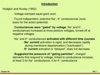
Hodgkin and Huxley. Taken from: http://icwww.epfl.ch/~gerstner/SPNM/node14.html. General Membrane Equation (a very important Equation, used everywhere!). Hodgkin Huxley Model:. charging current. Ion channels. with. and. Hodgkin Huxley Model:.

E N D
Hodgkin and Huxley Taken from: http://icwww.epfl.ch/~gerstner/SPNM/node14.html
General Membrane Equation (a very important Equation, used everywhere!) Hodgkin Huxley Model: charging current Ionchannels with and
Hodgkin Huxley Model: Introducing time-dependence so as to get an Action Potential modelled Following Hodgkin and Huxley (using rising AND falling functions): Resulting time-dependent Membrane Equation
40 20 V 0 m 20 V 40 60 80 0 5 10 15 20 t ms 40 20 V 0 m 20 V 40 60 80 0 5 10 15 20 t ms 40 20 V 0 m 20 V 40 60 80 0 5 10 15 20 t ms Hodgkin-Huxley Model: Action Potential / Threshold Iinj = 0.42 nA Short, weak current pulses depolarize the cell only a little. Iinj = 0.43 nA Iinj = 0.44 nA An action potential is elicited when crossing the threshold.
Hodgkin Huxley Model: asymptotic value • voltage dependent gating variables time constant with (u) (for the giant squid axon)
Derivative Solution: =
action potential • If u increases, m increases -> Na+ ions flow into the cell • at high u, Na+ conductance shuts off because of h • h reacts slower than m to the voltage increase • K+ conductance, determined by n, slowly increases with increased u
Hodgkin Huxley Model: Let’s see it in action! HHsim (seminar thema!)
Voltage clamp method • developed 1949 by Kenneth Cole • used in the 1950s by Alan Hodgkin and Andrew Huxley to measure ion current while maintaining specific membrane potentials
Large depolarization Voltage clamp method Small depolarization Ic: capacity current Il: leakage current
40 20 V 0 m 20 V 40 60 80 0 5 10 15 20 t ms 40 20 V 0 m 20 V 40 60 80 0 5 10 15 20 t ms 40 20 V 0 m 20 V 40 60 80 0 5 10 15 20 t ms Hodgkin-Huxley Model: Firing Latency Iinj = 0.45 nA Iinj = 0.65 nA A higher current reduces the time until an action potential is elicited. Iinj = 0.85 nA
40 20 V 0 m 20 V 40 60 80 0 5 10 15 20 t ms 40 20 V 0 m 20 V 40 60 80 0 5 10 15 20 t ms 40 20 V 0 m 20 V 40 60 80 0 5 10 15 20 t ms Hodgkin-Huxley Model: Firing Latency Iinj = 0.45 nA Iinj = 0.65 nA A higher current reduces the time until an action potential is elicited. Iinj = 0.85 nA
40 20 V 0 m 20 V 40 60 80 0 5 10 15 20 25 30 t ms 40 20 V 0 m 20 V 40 60 80 0 5 10 15 20 25 30 t ms 40 20 V 0 m 20 V 40 60 80 0 5 10 15 20 25 30 t ms Hodgkin-Huxley Model: Refractory Period Longer current pulses will lead to more action potentials. However, the next action potential can only occur after a “waiting period” during which the cell return to its normal state. This “waiting period” is called the refractory period. Iinj = 0.5 nA Iinj = 0.5 nA Iinj = 0.5 nA
40 20 V 0 m 20 V 40 60 80 0 20 40 60 80 100 t ms 40 20 V 0 m 20 V 40 60 80 0 20 40 60 80 100 t ms 40 20 V 0 m 20 V 40 60 80 0 20 40 60 80 100 t ms Hodgkin-Huxley Model: Firing Rate Iinj = 0.2 nA When injecting current for longer durations an increase in current strength will lead to an increase of the number of action potentials per time. Thus, the firing rate of the neuron increases. The maximum firing rate is limited by the absolute refractory period. Iinj = 0.3 nA Iinj = 0.6 nA
Varying firing properties Rhythmic burst in the absence of synaptic inputs ??? Influence of the neurotransmitter Acetylcholin Influence of steady hyperpolarization
Action Potential / Shapes: Cat - Heart Rat - Muscle Squid Giant Axon
Open channels per mm2 membrane area Local current loops Time Distance Propagation of an Action Potential: Action potentials propagate without being diminished (active process). Action potentials propagate without being diminished (active process). All sites along a nerve fiber will be depolarized until the potential passes threshold. As soon as this happens a new AP will be elicited at some distance to the old one. Action potentials propagate without being diminished (active process). All sites along a nerve fiber will be depolarized until the potential passes threshold. As soon as this happens a new AP will be elicited at some distance to the old one. Main current flow is across the fiber.
Structure of a Neuron: At the dendrite the incoming signals arrive (incoming currents) At the soma current are finally integrated. At the axon hillock action potential are generated if the potential crosses the membrane threshold The axon transmits (transports) the action potential to distant sites CNS Systems At the synapses are the outgoing signals transmitted onto the dendrites of the target neurons Areas Local Nets Neurons Synapses Molekules
Chemical synapse Neurotransmitter Receptors
Neurotransmitters Chemicals (amino acids, peptides, monoamines) that transmit, amplify and modulate signals between neuron and another cell. Cause either excitatory or inhibitory PSPs. Glutamate – excitatory transmitter GABA, glycine – inhibitory transmitter
Synaptic Transmission: Synapses are used to transmit signals from the axon of a source to the dendrite of a target neuron. Synapses are used to transmit signals from the axon of a source to the dendrite of a target neuron. There are electrical (rare) and chemical synapses (very common) Synapses are used to transmit signals from the axon of a source to the dendrite of a target neuron. There are electrical (rare) and chemical synapses (very common) At an electrical synapse we have direct electrical coupling (e.g., heart muscle cells). Synapses are used to transmit signals from the axon of a source to the dendrite of a target neuron. There are electrical (rare) and chemical synapses (very common) At an electrical synapse we have direct electrical coupling (e.g., heart muscle cells). At a chemical synapse a chemical substance (transmitter) is used to transport the signal. Synapses are used to transmit signals from the axon of a source to the dendrite of a target neuron. There are electrical (rare) and chemical synapses (very common) At an electrical synapse we have direct electrical coupling (e.g., heart muscle cells). At a chemical synapse a chemical substance (transmitter) is used to transport the signal. Electrical synapses operate bi-directional and are extremely fast, chem. syn. operate uni-directional and are slower. Synapses are used to transmit signals from the axon of a source to the dendrite of a target neuron. There are electrical (rare) and chemical synapses (very common) At an electrical synapse we have direct electrical coupling (e.g., heart muscle cells). At a chemical synapse a chemical substance (transmitter) is used to transport the signal. Electrical synapses operate bi-directional and are extremely fast, chem. syn. operate uni-directional and are slower. Chemical synapses can be excitatory or inhibitory they can enhance or reduce the signal change their synaptic strength (this is what happens during learning).
Axon Structure of a Chemical Synapse: Motor Endplate (Frog muscle) Synaptic cleft vesicles Active zone Muscle fiber Presynaptic membrane Postsynaptic membrane Synaptic cleft
Pre-synaptic action potential Concentration of transmitter in the synaptic cleft Post-synaptic action potential What happens at a chemical synapse during signal transmission: The pre-synaptic action potential depolarises the axon terminals and Ca2+-channels open. The pre-synaptic action potential depolarises the axon terminals and Ca2+-channels open. Ca2+ enters the pre-synaptic cell by which the transmitter vesicles are forced to open and release the transmitter. The pre-synaptic action potential depolarises the axon terminals and Ca2+-channels open. Ca2+ enters the pre-synaptic cell by which the transmitter vesicles are forced to open and release the transmitter. Thereby the concentration of transmitter increases in the synaptic cleft and transmitter diffuses to the postsynaptic membrane. The pre-synaptic action potential depolarises the axon terminals and Ca2+-channels open. Ca2+ enters the pre-synaptic cell by which the transmitter vesicles are forced to open and release the transmitter. Thereby the concentration of transmitter increases in the synaptic cleft and transmitter diffuses to the postsynaptic membrane. Transmitter sensitive channels at the postsyaptic membrane open. Na+ and Ca2+ enter, K+ leaves the cell. An excitatory postsynaptic current (EPSC) is thereby generated which leads to an excitatory postsynaptic potential (EPSP).
Neurotransmitters and their (main) Actions: Transmitter Channel-typ Ion-current Action Transmitter Channel-typ Ion-current Action Acetylecholin nicotin. Receptor Na+ and K+ excitatory Transmitter Channel-typ Ion-current Action Acetylecholin nicotin. Receptor Na+ and K+ excitatory Glutamate AMPA / Kainate Na+ and K+ excitatory Transmitter Channel-typ Ion-current Action Acetylecholin nicotin. Receptor Na+ and K+ excitatory Glutamate AMPA / Kainate Na+ and K+ excitatory GABA GABAA-Receptor Cl- inhibitory Transmitter Channel-typ Ion-current Action Acetylecholin nicotin. Receptor Na+ and K+ excitatory Glutamate AMPA / Kainate Na+ and K+ excitatory GABA GABAA-Receptor Cl- inhibitory Glycine Cl- inhibitory Transmitter Channel-typ Ion-current Action Acetylecholin nicotin. Receptor Na+ and K+ excitatory Glutamate AMPA / Kainate Na+ and K+ excitatory GABA GABAA-Receptor Cl- inhibitory Glycine Cl- inhibitory Acetylecholin muscarin. Rec. - metabotropic, Ca2+ Release Transmitter Channel-typ Ion-current Action Acetylecholin nicotin. Receptor Na+ and K+ excitatory Glutamate AMPA / Kainate Na+ and K+ excitatory GABA GABAA-Receptor Cl- inhibitory Glycine Cl- inhibitory Acetylecholin muscarin. Rec. - metabotropic, Ca2+ Release Glutamate NMDA Na+, K+, Ca2+ voltage dependent blocked at resting potential
Structure of a Neuron: At the dendrite the incoming signals arrive (incoming currents) At the soma current are finally integrated. At the axon hillock action potential are generated if the potential crosses the membrane threshold The axon transmits (transports) the action potential to distant sites CNS Systems At the synapses are the outgoing signals transmitted onto the dendrites of the target neurons Areas Local Nets Neurons Synapses Molekules
Chemical synapse Neurotransmitter Receptors
Neurotransmitters Chemicals (amino acids, peptides, monoamines) that transmit, amplify and modulate signals between neuron and another cell. Cause either excitatory or inhibitory PSPs. Glutamate – excitatory transmitter GABA, glycine – inhibitory transmitter
Synaptic Transmission: Synapses are used to transmit signals from the axon of a source to the dendrite of a target neuron. Synapses are used to transmit signals from the axon of a source to the dendrite of a target neuron. There are electrical (rare) and chemical synapses (very common) Synapses are used to transmit signals from the axon of a source to the dendrite of a target neuron. There are electrical (rare) and chemical synapses (very common) At an electrical synapse we have direct electrical coupling (e.g., heart muscle cells). Synapses are used to transmit signals from the axon of a source to the dendrite of a target neuron. There are electrical (rare) and chemical synapses (very common) At an electrical synapse we have direct electrical coupling (e.g., heart muscle cells). At a chemical synapse a chemical substance (transmitter) is used to transport the signal. Synapses are used to transmit signals from the axon of a source to the dendrite of a target neuron. There are electrical (rare) and chemical synapses (very common) At an electrical synapse we have direct electrical coupling (e.g., heart muscle cells). At a chemical synapse a chemical substance (transmitter) is used to transport the signal. Electrical synapses operate bi-directional and are extremely fast, chem. syn. operate uni-directional and are slower. Synapses are used to transmit signals from the axon of a source to the dendrite of a target neuron. There are electrical (rare) and chemical synapses (very common) At an electrical synapse we have direct electrical coupling (e.g., heart muscle cells). At a chemical synapse a chemical substance (transmitter) is used to transport the signal. Electrical synapses operate bi-directional and are extremely fast, chem. syn. operate uni-directional and are slower. Chemical synapses can be excitatory or inhibitory they can enhance or reduce the signal change their synaptic strength (this is what happens during learning).