Download
1 / 39
390 likes | 397 Views
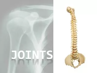
Articulations/Joints. Joint Classification A. Structural Two criteria: 1. +/- synovial cavity- space between bones often fluid-filled 2. Type of connective tissue binding bones Fibrous-no synovial cavity-bones held by fibrous connective tissue

E N D
Joint Classification A. Structural Two criteria: 1. +/- synovial cavity-space between bones often fluid-filled 2. Type of connective tissue binding bones Fibrous-no synovial cavity-bones held by fibrous connective tissue Cartilaginous-no synovial cavity-bones held by cartilage Synovial-bones held by connective tissue of the articular capsule and ligaments B. Functional Criteria: 1. Degree of movement Synarthrosis-an immovable joint Amphiarthrosis-a slightly movable joint Diarthrosis-a freely movable joint-synovial
Fibrous Joints Sutures-example of an immovable joint (synarthrosis) Syndesmoses-a fibrous joint that has greater distance between articulating bones than a suture (e.g. the tibiofibular joint, where the tibiofibular ligament conencts the tibia and fibula). Gomphoses-a fibrous joint where a cone-shaped peg fits into a socket- (e.g. articulation between the teeth roots and alveoli of the alveolar processes of the maxillae)
Cartilaginous Joints Synchondroses-a joint in which the connecting material is hayline cartilage (e.g. the epiphyseal plate connecting the epiphysis and diaphysis of a growing bone) Symphyses-a cartilaginous joint in which the ends of the articulating bone are covered with hyaline cartilage. Occurs at the midline. (e.g. the pubic symphysis connects the hip bones)
Synovial Joints • Structure of Synovial Joints • Unique in that they have a synovial cavity between articulating bones. • Classified as diarthroses • Bones are covered by a layer of hyaline cartilage called articular cartilage • ArticularCapsule-asleeve-like covering around a synovial joint consists • of two layers: an outer fibrous capsule and an inner synovial membrane. • The fibrous capsule (mostly collagen fibers)attaches to the periosteum of • the articulating bones. Some fibrous capsules are arranged in bundles • called ligaments. • The synovial membrane (areolar connective tissue with elastic fibers) • may include accumulation of fat pads called articular fat pads. • SynovialFluid-Secreted by synovial membranes (hayluronic acid and • interstitial fluid). Lubricates joints, absorbes shocks, supplies nutrients • and removes wastes from chondrocytes within articular cartilage. • Accessory Ligaments- Extracapsular ligaments (outside the capsule). • Articular discs or menisci- stability, better joint fit, lubrication
Synovial Joints (cont’d) Torn Cartilage-tearing of articular disks (menisci). Sprain-forcible stretching of a joint that stretches or tears ligaments without dislocating bones. Strain-stretched or partially torn muscle. Bursae- Sac-like strutures that relieve friction in some joints (shoulder and knee). Not part of synovial joints but the capsules consist of connective tissue lined with synovial membrane. Tendon sheaths are tube-like bursae that wrap some tendons to lessen friction. Bursitis is an inflammation of bursae.
Types of joint movement at synovial joints Gliding-bones move side-to-side, no significant alteration in angle between bones
Types of joint movement at synovial joints-angular Flexion-a decrease in the angle between articulating bones Extension-and increase in the angle between articulating bones Hyperextension-continuation of extension beyond the anatomical position
Types of joint movement at synovial joints-angular Flexion-a decrease in the angle between articulating bones Extension-and increase in the angle between articulating bones Hyperextension-continuation of extension beyond the anatomical position
Types of joint movement at synovial joints-angular Flexion-a decrease in the angle between articulating bones Extension-and increase in the angle between articulating bones Hyperextension-continuation of extension beyond the anatomical position
Types of joint movement at synovial joints-angular Lateral Flexion-decrease in the angle between articulating bones
Types of joint movement at synovial joints-angular Abduction-movement away from the midline Adduction-movement toward the midline Circumduction-movement of the distal end of a body part in a circle
Types of joint movement at synovial joints-angular Abduction-movement away from the midline Adduction-movement toward the midline
Types of joint movement at synovial joints-angular Circumduction-movement of the distal end of a body part in a circle
Types of Movements at Synovial Joints Rotation-a bone revolving around its own longitudinal axis e.g. turning the head from side-to-side at the atlanto-axial joint (between the atlas and axis) when shaking your head-”no”. In limbs the rotation is defined relative to the midline e.g. medial or internal rotation of the humerus Special Movements-only occur at certain joints. 1. Elevation-upward movement opposes depression 2. Protraction-anterior movement opposes retraction 3. Inversion-inward movement of feet opposes eversion 4. Dorsiflexion-bending the foot at the ankle (standing on heels) opposes plantar flexion 5. Supination-radioulnar movement where palm is face-up opposes pronation (palm is face down) 6. Opposition- movement ofthumb to touch fingers
Tendons and Ligaments A tendon is a fibrous connective tissue which attaches muscle to bone. Tendons may also attach muscles to structures such as the eyeball. A tendon serves to move the bone or structure. A ligament is a fibrous connective tissue which attaches bone to bone, and usually serves to hold structures together and keep them stable.
Tendons and Ligaments The ligament or tendon is split into smaller entities called fascicles. The fascicle contains the basic fibril of the ligament or tendon, and the fibroblasts, which are the biological cells that produce the ligament or tendon. There is a structural characteristic at this level that plays a significant role in the mechanics of ligaments and tendons: the crimp of the fibril. The crimp is the waviness of the fibril.
Six Types of Synovial Joints-Planar Articulating surfaces are flat or slightly curved-primarily Involved in gliding movements
Six Types of Synovial Joints-Hinge The convex surface of one bone fits into the concave surface of another-monoaxial (movement in one axis)
Six Types of Synovial Joints-Pivot The rounded or pointed surface of one bone articulates with a ring formed by a bone and a ligament-monoaxial
Six Types of Synovial Joints-Condyloid The oval-shaped convexity of one bone fits into the oval-shaped depression in another bone-biaxial
Six Types of Synovial Joints-Saddle An articular surface of one bone fits into the saddle-shaped surface of another bone-biaxial
Six Types of Synovial Joints-Ball-and Socket The ball-like surface of one bone fits into a cup-like depression of another bone
Factors that affect range-of-motion at synovial joints Structure and shape of articulating bones Strength and tension of joint ligaments Arrangement and tension of muscles Contact of soft body parts Hormones- e.g. relaxin relaxes symphasis pubis Disuse- e.g. lack of ROM after immobilization
Humeroscapular (Glenohumeral) Joint Rotator cuff injury
Joint disorders and aging Aging-decreased synovial fluid, thinner articular cartilage, ligaments shorten. Nearly everyone develops degeneration in Knees, Elbows, Hips, and Shoulders. Arthroplasty-repair of degenerated joints with artificial parts e.g. full or partial hip replacement (head of the femur +/- the acetabulum. Disorders- a. Rheumatism/arthritis-inflamation of bones, ligaments, tendons, or muscles not caused by direct injury. b. Osteoarthritis-degenerative disease resulting in lost joint cartilage c. Rheumatoid arthritis-autoimmune disease attacking joint linings and cartilage d. Gouty arthritis-uric acid build up resulting in crystals deposited in joints-especially feet-can destroy joint-causing fusion e. Lyme Disease-bacterial infection via tick bite affects large joints (knee, ankle, elbow, hip, wrist)