Download
1 / 1
10 likes | 107 Views
NSF NIRT-0507083: An Optimized Nanosphere Platform for High Resolution Multi-Modality Imaging Applications. Acknowledgments. Other Funding:

E N D
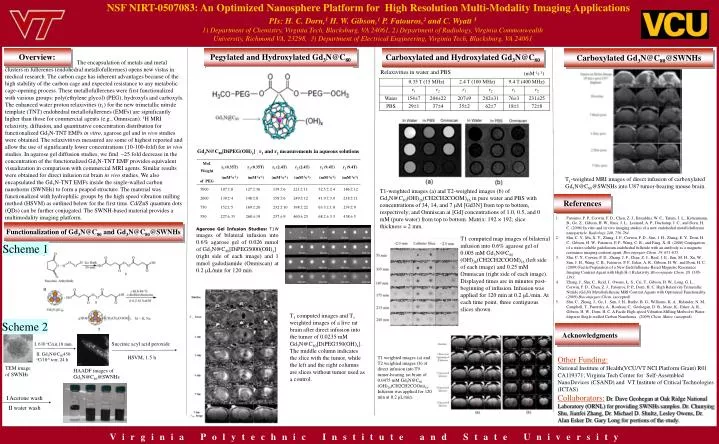
NSF NIRT-0507083: An Optimized Nanosphere Platform for High Resolution Multi-Modality Imaging Applications Acknowledgments Other Funding: National Institute of Health(VCU/VT NCI Platform Grant) R01 CA119371; Virginia Tech Center for Self-Assembled NanoDevices (CSAND) and VT Institute of Critical Technologies (ICTAS) Collaborators:Dr. Dave Geohegan at Oak Ridge National Laboratory (ORNL) for providing SWNHs samples. Dr. Chunying Shu, Jianfei Zhang, Dr. Michael D. Shultz, Lesley Owens, Dr. Alan Esker Dr. Gary Long for portions of the study. PIs: H. C. Dorn,1 H. W. Gibson,1 P. Fatouros,2 and C. Wyatt 31) Department of Chemistry, Virginia Tech, Blacksburg, VA 24061, 2) Department of Radiology, Virginia Commonwealth University, Richmond VA, 23298, 3) Department of Electrical Engineering, Virginia Tech, Blacksburg, VA 24061 Overview: Pegylated and Hydroxylated Gd3N@C80 Carboxylated and Hydroxylated Gd3N@C80 Carboxylated Gd3N@C80@SWNHs The encapsulation of metals and metal clusters in fullerenes (endohedral metallofullerenes) opens new vistas in medical research. The carbon cage has inherent advantages because of the high stability of the carbon cage and expected resistance to any metabolic cage-opening process. These metallofullerenes were first functionalized with various groups: poly(ethylene glycol) (PEG), hydroxyls and carboxyls. The enhanced water proton relaxivities (r1) for the new trimetallic nitride template (TNT) endohedral metallofullerenes (EMFs) are significantly higher than those for commercial agents (e.g., Omniscan). 1H MRI relaxivity, diffusion, and quantitative concentration distribution for functionalized Gd3N-TNT EMFs in vitro, agarose gel and in vivo studies were obtained. The relaxivitives measured are some of highest reported and allow the use of significantly lower concentrations (10-100-fold) for in vivo studies. In agarose gel diffusion studies, we find ~25 fold decrease in the concentration of the functionalized Gd3N-TNT EMF provides equivalent visualization in comparison with commercial MRI agents. Similar results were obtained for direct infusion rat brain in vivo studies. We also encapsulated the Gd3N-TNT EMFs inside the single-walled carbon nanohorns (SWNHs) to form a peapod structure. The material was functionalized with hydrophilic groups by the high speed vibration milling method (HSVM) as outlined below for the first time. Cd/ZnS quantum dots (QDs) can be further conjugated. The SWNH-based material provides a multimodality imaging platform. Relaxivities in water and PBS (mM-1s-1) Gd3N@C80[DiPEG(OH)x] : r1 and r2 measurements in aqueous solutions T1-weighted MRI images of direct infusion of carboxylated Gd3N@C80@SWNHs into U87 tumor-bearing mouse brain. T1-weighted images (a) and T2-weighted images (b) of Gd3N@C80(OH)26(CH2CH2COOM)16 in pure water and PBS with concentrations of 34, 14, and 7 μM [Gd3N] from top to bottom, respectively; and Omniscan at [Gd] concentrations of 1.0, 0.5, and 0 mM (pure water) from top to bottom. Matrix: 192 × 192; slice thickness = 2 mm. References • Fatouros, P. P., Corwin, F. D., Chen, Z. J., Broaddus, W. C., Tatum, J. L., Kettenmann, B., Ge, Z., Gibson, H. W., Russ, J. L., Leonard, A. P., Duchamp, J. C., and Dorn, H. C. (2006) In vitro and in vivo imaging studies of a new endohedral metallofullerene nanoparticle. Radiology 240, 756-764. • Shu, C. Y., Ma, X. Y., Zhang, J. F., Corwin, F. D., Sim, J. H., Zhang, E. Y., Dorn, H. C., Gibson, H. W., Fatouros, P. P., Wang, C. R., and Fang, X. H. (2008) Conjugation of a water-soluble gadolinium endohedral fulleride with an antibody as a magnetic resonance imaging contrast agent. Bioconjugate Chem. 19, 651-655. • Shu, C. Y., Corwin, F. D., Zhang, J. F., Chen, Z. J., Reid, J. E., Sun, M. H., Xu, W., Sim, J. H., Wang, C. R., Fatouros, P. P., Esker, A. R., Gibson, H. W., and Dorn, H. C. (2009) Facile Preparation of a New Gadofullerene-Based Magnetic Resonance Imaging Contrast Agent with High H-1 Relaxivity. Bioconjugate Chem. 20, 1186-1193. • Zhang, J., Shu, C., Reid, J., Owens, L. S., Cai, T., Gibson, H. W., Long, G. L., Corwin, F. D., Chen, Z. J., Fatouros, P. P., Dorn, H. C. High Relaxivity Trimetallic Nitride (Gd3N) Metallofullerene MRI Contrast Agents with Optimized Functionality. (2009) Bioconjugate Chem. (accepted) • Shu, C., Zhang, J., Ge, J., Sim, J. H., Burke, B. G., Williams, K. A., Rylander, N. M., Campbell, T., Puretzky, A., Rouleau, C., Geohegan, D. B., More, K., Esker, A. R., Gibson, H. W., Dorn, H. C. A Facile High-speed Vibration Milling Method to Water-disperse Single-walled Carbon Nanohorns . (2009) Chem. Mater. (accepted) Functionalization of Gd3N@C80 and Gd3N@C80@SWNHs Agarose Gel Infusion Studies: T1W images of bilateral infusion into 0.6% agarose gel of 0.026 mmol of Gd3N@C80[DiPEG5000(OH)x] (right side of each image) and 1 mmolgadodiamide (Omniscan) at 0.2 L/min for 120 min. T1 computed map images of bilaterial infusion into 0.6% agarose gel of 0.005 mM Gd3N@C80 (OH)26(CH2CH2COOM)16 (left side of each image) and 0.25 mM Omniscan (right side of each image). Displayed times are in minutes post-beginning of infusion. Infusion was applied for 120 min at 0.2 μL/min. At each time point, three contiguous slices shown. Scheme 1 T1 computed images and T1 weighted images of a live rat brain after direct infusion into the tumor of 0.0235 mM Gd3N@C80[DiPEG350(OH)x]. The middle column indicates the slice with the tumor, while the left and the right columns are slices without tumor used as a control. Scheme 2 Succinic acyl acid peroxide I. 630 oC/air,10 min. II. Gd3N@C80450 oC/10-6 torr, 24 h HSVM, 1.5 h T1 weighted images (a) and T2 weighted images (b) of direct infusion into T9 tumor-bearing rat brain of 0.0475 mM Gd3N@C80 (OH)26(CH2CH2COOm)16. Infusion was applied for 120 min at 0.2 μL/min. TEM image of SWNHs HAADF images of Gd3N@C80@SWNHs I Acetone wash II water wash Virginia Polytechnic Institute and State University