Download
1 / 1
20 likes | 196 Views
primary tumor. Cortical actin. F-actin. cortical actin. blood vessel. stress fibers. G-actin. lamellipodia and filopodia. Stress fiber. focal adhesion. secondary tumor. Introduction. Methods (continued). Results (continued).

E N D
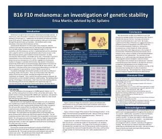
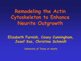
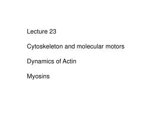
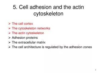
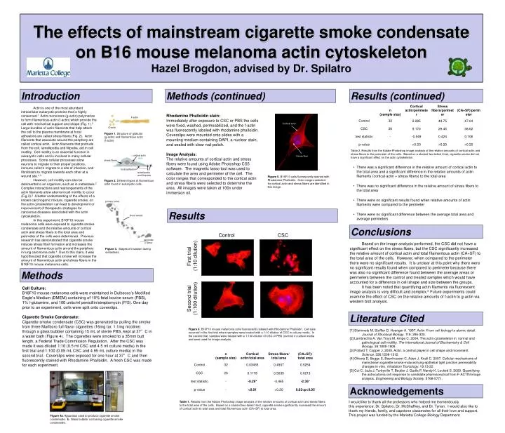
primary tumor Cortical actin F-actin cortical actin blood vessel stress fibers G-actin lamellipodia and filopodia Stress fiber focal adhesion secondary tumor Introduction Methods (continued) Results (continued) Actin is one of the most abundant intracellular eukaryotic proteins that is highly conserved.1 Actin monomers (g-actin) polymerize to form filamentous actin (f-actin) which provide the cell with mechanical support and shape (Fig. 1).2 Large bundles of actin filaments that help attach the cell to the plasma membrane at focal adhesions are called stress fibers (Fig. 2). Actin filaments that associate around the periphery are called cortical actin. Actin filaments that protrude from the cell, lamellipodia and filipodia, aid in cell motility. Cell motility is an essential function in eukaryotic cells and is involved in many cellular processes. Some cellular processes allow neurons to migrate to their proper positions, immune cells to migrate to a site of infection, and fibroblasts to migrate towards each other at a wound site.2,3 However, cell motility can also be detrimental to an organism, such as in metastasis.2 Complex interactions and rearrangements of the actin filaments allow aberrant cell motility to occur (Fig 3).2 A better understanding of the effects of a known carcinogenic mixture, cigarette smoke, on the actin cytoskeleton can lead to development or improvement of therapeutic strategies for cancerous diseases associated with the actin cytoskeleton. In this experiment, B16F10 mouse melanoma cells were exposed to cigarette smoke condensate and the relative amounts of cortical actin and stress fibers to the total area and perimeter of the cells were determined. Previous research has demonstrated that cigarette smoke induces stress fiber formation and increases the amount of filamentous actin around the periphery in lung carcinoma cells.4 Due to this claim, it was hypothesized that cigarette smoke will increase the amount of filamentous actin and stress fibers in the B16F10 mouse melanoma cells. Control CSC Rhodamine Phalloidin stain: Immediately after exposure to CSC or PBS the cells were fixed, washed, permeabilized, and the f-actin was fluorescently labeled with rhodamine phalloidin. Coverslips were mounted onto slides with a mounting medium containing DAPI, a nuclear stain, and sealed with clear nail polish. Image Analysis: The relative amounts of cortical actin and stress fibers were found using Adobe Photoshop CS5 software. The magnetic lasso tool was used to calculate the area and perimeter of the cell. The color ranges that corresponded to the cortical actin and stress fibers were selected to determine the area. All images were taken at 100x under immersion oil. First trial (1:10 dilution) Figure 1. Structure of globular (g-actin) and filamentious actin (f-actin). The effects of mainstream cigarette smoke condensate on B16 mouse melanoma actin cytoskeletonHazel Brogdon, advised by Dr. Spilatro Table 2. Results from the Adobe Photoshop image analysis of the relative amounts of cortical actin and stress fibers to the perimeter of the cells. Based on a student two-tailed t-test, cigarette smoke did not have a significant effect on the actin cytoskeleton. • There was a significant difference in the relative amount of cortical actin to the total area and a significant difference in the relative amounts of actin filaments (cortical actin + stress fibers) to the total area • There was no significant difference in the relative amount of stress fibers to the total area • There were no significant results found when relative amounts of actin filaments were compared to the perimeter • There were no significant difference between the average total area and average perimeters Second trial (1:100 dilution) Figure 5. B16F10 cells fluorescently stained with Rhodamine Phalloidin. Color ranges selected for cortical actin and stress fibers are identified in this image. Figure 2. Different types of filamentous actin found in eukaryotic cells. Results Conclusions Based on the image analysis performed, the CSC did not have a significant effect on the stress fibers, but the CSC significantly increased the relative amount of cortical actin and total filamentous actin (CA+SF) to the total area of the cells. However, when compared to the perimeter there were no significant results. It is unclear at this point why there were no significant results found when compared to perimeter because there was also no significant difference found between the average areas or perimeters between the control and treated samples which would have accounted for a difference in cell shape and size between the groups. It has been noted that quantifying actin filaments via fluorescent image analysis is very difficult and complex.5 Future experiments could examine the effect of CSC on the relative amounts of f-actin to g-actin via western blot analysis. Figure 3. Stages of invasion during metastasis. Methods • Cell Culture: • B16F10 mouse melanoma cells were maintained in Dulbecco’s Modified Eagle’s Medium (DMEM) containing of 10% fetal bovine serum (FBS), 1% l-glutamine, and 100 units/ml penicillin/streptomycin (P/S). One day prior to an experiment, cells were split onto coverslips. • Cigarette Smoke Condensate: • Cigarette smoke condensate (CSC) was generated by pulling the smoke from three Marlboro full flavor cigarettes (16mg tar, 1.1mg nicotine) through a glass bubbler containing 15 mL of sterile PBS, kept at 37°C in a water bath (Figure 4). The cigarettes were smoked to a 35mm butt length, a Federal Trade Commission Regulation. After the CSC was made it was diluted 1:10 (0.5 ml CSC and 4.5 ml culture media) in the first trial and 1:100 (0.05 mL CSC and 4.95 mL culture media) in the second trial. Coverslips were exposed for one hour at 37°C and then fluorescently stained with Rhodamine Phalloidin. A fresh CSC was made for each experiment. a b Literature Cited Figure 6. B16F10 mouse melanoma cells fluorescently labeled with Rhodamine Phalloidin. Cell lysis occurred in the first trial where samples were treated with a 1:10 dilution of CSC in culture media. In the second trial, samples were treated with a 1:100 dilution of CSC or PBS (control) in culture media and were used for image analysis. [1] Steinmets M, Stoffler D, Hoenger A. 1997. Actin: From cell biology to atomic detail. Journal of Structural Biology. 119: 295-320. [2] Lambrechts A, Van Troys M, Ampe C. 2004. The actin cytoskeleton in normal and pathological cell motility. The International Journal of Biochemistry & Cell Biology. 36:1809-1909. [3] Pollard T, Copper J. 2009. Actin, a central player in cell shape and movement. Science. 326:1208-1212. [4] Olivera D, Boggs S, Beenhouwer C, Aden J, Knall C. 2007. Cellular mechanisms of mainstream cigarette smoke-induced lung epithelial tight junction permeability changes in vitro. Inhalation Toxicology. 19:13-22 [5] Cui C, JaJa J, Turbyville T, Beutler J, Gudla P, Nandy K, Lockett S. 2009. Quantifying the astrocytoma cell response to candidate pharmaceutical from F-ACTIN image analysis. Engineering and Biology Society. 5768-5771. . Figure 4a. Apparatus used to produce cigarette smoke condensate. b. Glass bubbler containing cigarette smoke condensate. Acknowledgements I would like to thank all the professors who helped me tremendously throughout this experience: Dr. Spilatro, Dr. McShaffrey, and Dr. Tynan. I would also like to thank my friends, family, and capstone classmates for all their love and support. This project was funded by the Marietta College Biology Department. Table 1. Results from the Adobe Photoshop image analysis of the relative amounts of cortical actin and stress fibers to the total area of the cells. Based on a student two-tailed t-test, cigarette smoke significantly increased the amount of cortical actin to total area and total filamentous actin (CA+SF) to total area.