Download
1 / 86
890 likes | 1.13k Views
ENDOCRINE SYSTEM. PHYSIOLOGY. Endocrine vs Nervous System. NERVOUS. ENDOCRINE. Uses chemical hormones released from glands into the blood. Uses action potentials along axons and chemical neurotransmitters at synapses. Receptors are on the plasma membranes of target cells or intercellular.

E N D
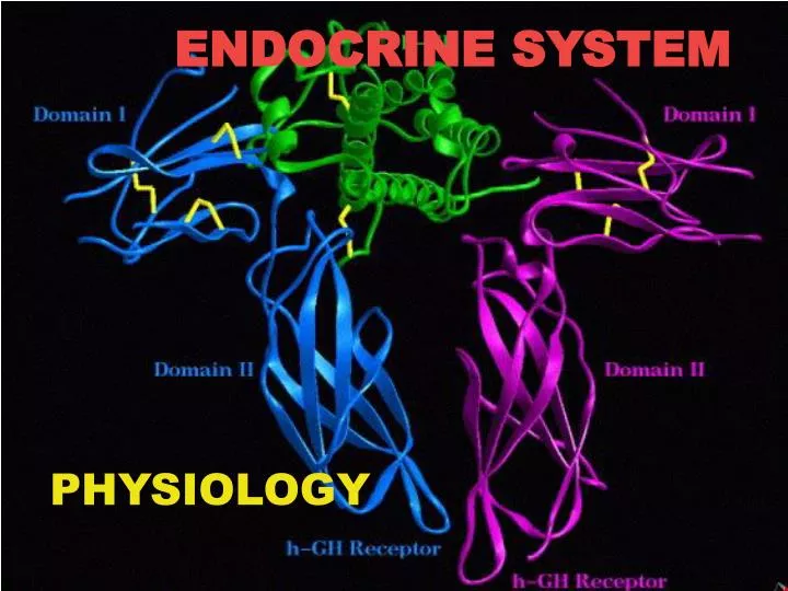
ENDOCRINE SYSTEM PHYSIOLOGY
Endocrine vs Nervous System NERVOUS ENDOCRINE • Uses chemical hormones released from glands into the blood • Uses action potentials along axons and chemical neurotransmitters at synapses • Receptors are on the plasma membranes of target cells or intercellular • Receptors are on post-synaptic membrane • Signals are very fast (milliseconds) • Signals are slower (seconds to days) • Response is immediate but short-lived • Response is delayed but more sustained
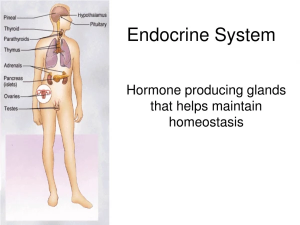
Fig. 11.1 P. 286 Endocrine glands are ductless glands
Blurring the edges Specialized neurons can secrete chemicals into the blood rather than synaptic cleft. • Chemical secreted is called neurohormone. • Hypothalamus primary secretor of neurohormones. • Some chemicals like norepinephrine is • both a neurotransmitter and a hormone.
Characteristics of Hormones • Hormones: • exert their effects some distance from where they are produced • are active under very low (picogram to nanogram) concentrations in the blood • usually have a short half-life in the body - several seconds to 60 mins. They are degraded by enzymes in their target cells or in the kidney or liver.
Characteristics of Hormones • Hormones bring about their effects by altering cell activity. The precise response depends on the target cell type. Typical cellular effects include: • Altering plasma membrane permeability • Stimulating protein synthesis • Activating enzymes • Inducing secretory activity • Stimulating mitosis
Characteristics of Hormones • Hormones levels depend on: • Rate of release • Speed of inactivation and removal from the body • Pharmacological levels of a hormone may have different functions than physiological levels of the hormone
Chemical Classes of Hormones • Amines - Derived from tyrosine or tryptophan.Includes: epinephrine, T4, and melatonin. • Proteins and peptides - Made from amino acid chains. Includes: antidiuretic hormone, growth hormone, and insulin. • Glycoproteins - A polypeptide chain bound to one or more carbohydrates.Includes: follicle-stimulating hormone and luteinizing hormone. • Steroids - Lipids derived from cholesterol.Includes: testosterone, estradiol, and cortisol.
Chemical Classes of Hormones • Hormones can also be divided into: • Polar: • H20 soluble. • Catecholamines, peptides, and glycoproteins • Nonpolar (lipophilic): • H20 insoluble (but lipid soluble). • Can gain entry into target cells • Steroid hormones and T4 • Pineal gland secretes melatonin: • Has properties of both H20 soluble and lipophilic hormones.
Control of Hormone Release • Synthesis and release of most hormones are regulated by a negative feedback system. As hormone levels rise, they cause target organ effects which inhibit further hormone release.
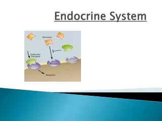
Hormone - Target Cell Specificity • Hormones circulate to virtually all tissues but influence the activity of only certain tissue cells, known as its target cells.
Hormone - Target Cell Specificity • Hormone-receptor interaction depends upon three factors: • Blood levels of the hormone • Relative number of receptors for that hormone on the target cell • Affinity of the bond between the hormone and the receptor
Hormone - Target Cell Specificity • Receptors are dynamic structures: they can respond to rising levels of hormones by increasing in number (up-regulation) or respond to prolonged exposure to high hormone concentrations by reducing the number of receptors (down-regulation).
Mechanisms of Hormone Action • Hormones: • Diffuse through the cell membrane and bind to intracellular receptors (steroid hormones & T4) or bind to receptors on the membrane of distant cells (amino-acid based hormones). • Carry out their effects by direct gene activation (steroids) or through signal transduction systems (amino-acid based).
Lipophilic steroid and thyroid hormones are attached to plasma carrier proteins. • Hormones dissociate from carrier proteins to pass through lipid component of the target plasma membrane. • Receptors for the lipophilic hormones are known as nuclear hormone receptors. • Steroid receptors function within cell to activate gene transcription. Fig. 11.4 P. 292
Each nuclear hormone receptor has 2 regions: • A ligand (hormone)-binding domain. • DNA-binding domain. • Receptor must be activated by binding to hormone before binding to specific region of DNA called HRE (hormone responsive element). • Located adjacent to gene that will be transcribed. Fig. 11.5 P. 293
Fig. 11.3 P. 289
The carrier protein for T4 is thyroxine-binding globulin (TBG). • Free T4 passes into cytoplasm and is converted to T3. • Nonspecific binding proteins shuttle it to the nucleus • Receptor proteins are in the nucleus. Fig. 11.6 P. 294
T3 binds to ligand-binding domain. • Other half-site is vitamin A derivative (9-cis-retinoic acid). • DNA-binding domain can then bind to the half-site of the HRE. • Two partners can bind to the DNA to activate HRE. • Stimulate transcription of genes. Fig. 11.7 P. 294
Adenylate Cyclase-cAMP Fig. 11.8 P. 295
Polypeptide or glycoprotein hormones bind to receptor protein causing dissociation of a subunit of G-protein.
G-protein subunit binds to and activates adenylate cyclase. ATP cAMP + PPi
cAMP attaches to inhibitory subunit of protein kinase. Inhibitory subunit dissociates and protein kinase is activated.
Phosphorylates enzymes within the cell to produce hormone’s effects.
cAMP inactivated by phosphodiesterase, which hydrolyzes cAMP to inactive fragments.
Phospholipase-C-Ca2+ Fig. 11.9 P. 297
Phospholipase-C-Ca2+ • Ca2+ diffuses into the cytoplasm and binds to calmodulin. • Calmodulin activates specific protein kinase enzymes.
Epi Can Act Through Two 2nd Messenger Systems Fig. 11.10 P. 297
Tyrosine Kinase • Stimulate glycogen, fat and protein synthesis. • Stimulate insertion of GLUT-4 carrier proteins. Fig. 11.11 P. 298
CONTROL OF RELEASE TARGET ORGAN HORMONE GLAND EFFECTS OF HYPER- AND HYPOSECRETION NORMAL EFFECTS OF HORMONE
Pituitary • Pituitary gland (hypophysis) is located in the diencephalon. • Structurally and functionally divided into: • Anterior lobe = Adenohypophysis • Posterior lobe = Neurohypophysis Fig. 11.12 P. 299
Pituitary • Anterior pituitary (adenohypophysis): • Master gland • Derived from a pouch of epithelial tissue that migrates upward from the mouth. • Consists of 2 main parts: • Pars distalis: anterior portion. • Pars tuberalis: thin extension in contact with the infundibulum. • Posterior pituitary(neurohypophysis): • Formed by downgrowth of the brain during fetal development. • Is in contact with the infundibulum.
Pituitary Hormones Fig. 11.14 P. 302
Gonadotropins • The gonadotropins are: • Follicle-stimulating hormone (FSH) - Responsible for gamete production in both sexes. • Luteinizing Hormone (LH) - In females works with FSH to cause follicle development, and then independently is responsible for ovulation. In males it is sometimes called interstitial cell-stimulating hormone (ICSH), because itstimulates the interstitial cells to produce testosterone
THYROID-STIMULATING HORMONE • Thyroid-stimulating Hormone (Thyrotropin; TSH) - chain of 96 amino acids; chain of 112 amino acids. • Acts on the thyroid follicle cells to stimulate thyroid hormone synthesis
ADRENOCORTICOTROPIC HORMONE • Adrenocorticotropic Hormone (ACTH) - polypeptide of 39 amino acids • Stimulates cells of adrenal cortex to increase steroid synthesis and secretion
GROWTH HORMONE • Growth Hormone (Somatotropin) - protein of 191 amino acids. • General anabolic stimulant • Works by stimulating production of an insulin-like growth factor (IGF-1; somatomedin C) in the liver • IGF-1 stimulates uptake of amino acids and sulfur, particularly on developing bone, and mobilizes fat from fat depots
GROWTH HORMONE • Gigantism refers to a condition characterized by extreme physical size and stature due to a hypersecretion of growth hormone during infancy, childhood or adolescence 12 year-old with mother
GROWTH HORMONE • Dwarfism results from a GH deficiency in childhood, leading to a maximum height of 4 feet typically with normal body proportions. If diagnosed before puberty, hormone replacement therapy can promote nearly normal growth. Dwarfed brothers with researcher in India
PROLACTIN • Prolactin (PRL) - Protein hormone of 199 amino acids.In females it stimulates milk production by the mammary glands. There is some evidence it enhances testosterone production in males. • Release is inhibitedin non-pregnant women. As estrogen and progesterone levels rise late in pregnancy, it stimulates prolactin release.
PROLACTIN • Hyperprolactinaemia can cause menstrual problems in females and breast enlargement in males. • Pituitary tumors is a major cause of the condition.
MELANOCYTE-STIMULATING HORMONE • Melanocyte-stimulating Hormone (MSH) - Derived from a prohormone called pro-opiomelanocortin (POMC) - chain of 13 amino acids; chain of 18 amino acids; chain of 12 amino acids. The major products of POMC is -endorphins, MSH, and ACTH.
MELANOCYTE-STIMULATING HORMONE • Stimulates pigmentation in fishes, amphibians and reptiles by enhancing the dispersion of melanin from melanocytes • In birds and mammals, blood levels are insignificant. It will cause darkening of the skin if injected into the circulation, but may be more important as a neurotransmitter in humans than in skin pigmentation.
NEUROHYPOPHYSIS • Hypothalamus neuron cell bodies produce: • ADH: supraoptic nuclei. • Oxytocin: paraventricular nuclei. • Transported along the hypothalamo-hypophyseal tract. • Stored and released from posterior pituitary. Fig. 11.13 P. 301
ANTIDIURETIC HORMONE • Antidiuretic Hormone (ADH; vasopressin) - oligopeptide of 9 amino acids. • The main regulator of body fluid osmolarity • Increases the reabsorption rate of water in kidney tubule cells; under high concentrations promotes vasoconstriction • Secretion is regulated in the hypothalamus by osmoreceptors, which sense water concentration
OXYTOCIN • Oxytocin - oligopeptide of 9 amino acids • hormonal trigger for milk ejection (the letdown reflex) in women whose breasts are actively producing milk • a strong stimulant of uterine contraction, and is released in progressively greater amounts as birth nears.
Hypothalamic Control of the Anterior Pituitary • Hormonal control rather than neural. • Hypothalamus neurons synthesize releasing and inhibiting hormones. • Hormones secreted into the hypothalamo-hypophyseal portal system regulate the secretions of the anterior pituitary Fig. 11.15 P. 303
Secretions are controlled by negative feedback inhibition by target gland hormones. • Negative feedback at 2 levels: • The target gland hormone can act on the hypothalamus and inhibit secretion of releasing hormones. • The target gland hormone can act on the anterior pituitary and inhibit response to the releasing hormone. Fig. 11.17 P. 304
Adrenal Gland • Paired organs that cap the kidneys. • Each gland consists of an outer cortex and inner medulla.
Adrenal Cortex • Adrenal cortex: • Does not receive neural innervation. • Must be stimulated hormonally (ACTH). • Consists of 3 zones: • Zona glomerulosa. • Zona fasciculata. • Zona reticularis. • Secretes corticosteroids. Fig. 11.18 P. 305