Download
1 / 41
410 likes | 514 Views
Chapter 16: The Knee and Related Structures. Complex joint that endures great amounts of trauma due to extreme amounts of stress that are regularly applied Hinge joint w/ a rotational component Stability is due primarily to ligaments, joint capsule and muscles surrounding the joint

E N D
Complex joint that endures great amounts of trauma due to extreme amounts of stress that are regularly applied • Hinge joint w/ a rotational component • Stability is due primarily to ligaments, joint capsule and muscles surrounding the joint • Designed for stability w/ weight bearing and mobility in locomotion
Prevention of Knee Injuries • Physical Conditioning and Rehabilitation • Total body conditioning is required • Strength, flexibility, cardiovascular and muscular endurance, agility, speed and balance • Muscles around the hip and knee must be conditioned to maximize stability • Shoe Type • Reduction in injuries • Use of cleats that allow for foot control but that do not fix foot to the ground
Functional and Prophylactic Knee Braces • Used to prevent and reduce severity of knee injuries • Provide degree of support to unstable knee • Can be custom molded and designed to control rotational forces and tibial translation
Assessing the Knee Joint • Determining the mechanism of injury is critical • History- Current Injury • Past history • Mechanism- what position was your body in? • Did the knee collapse? • Did you hear or feel anything? • Could you move your knee immediately after injury or was it locked? • Did swelling occur? • Where was the pain
History - Recurrent or Chronic Injury • What is your major complaint? • When did you first notice the condition? • Is there recurrent swelling? • Does the knee lock or catch? • Is there severe pain? • Grinding or grating? • Does it ever feel like giving way? • What does it feel like when ascending and descending stairs? • What past treatment have you undergone? • Any training or footwear changes?
Observation • Walking, half squatting, going up and down stairs • Swelling, ecchymosis • Assessment of muscle symmetry/atrophy • What is the athlete’s level of function? • Does the athlete limp? • Full weight bearing? • Does athlete exhibit normal knee mechanics during function?
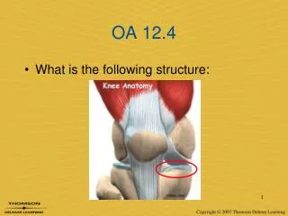
Palpation • Athlete should be supine or sitting at edge of table with knee flexed to 90 degrees • Should assess bony structures checking for bony deformity and/or pain • Soft tissue • Lateral ligaments • Joint line • Assess for pain and tenderness • Menisci
Special Tests for Knee Instability • Use endpoint feel to determine stability • Classification of Joint Instability • Knee laxity includes both straight and rotary instability • Translation (tibial translation) refers to the glide of tibial plateau relative to the femoral condyles • As the damage to stabilization structures increases, laxity and translation also increase • Valgus and Varus Stress Tests • Used to assess the integrity of the MCL and LCL respectively
Valgus Stress Test Varus Stress Test
Lachman Drawer Test • Will not force knee into painful flexion immediately after injury • Reduces hamstring involvement • At 30 degrees of flexion an attempt is made to translate the tibia anteriorly on the femur • A positive test indicates damage to the ACL
Apley’s Compression Test • Hard downward pressure is applied w/ rotation • Pain indicates a meniscal injury • Used to detect meniscus tear
Recognition and Management of Specific Injuries • Medial Collateral Ligament Sprain • Cause of Injury • Result of severe blow or outward twist – valgus force • Signs of Injury - Grade I • Little fiber tearing or stretching • Stable valgus test • Little or no joint effusion • Some joint stiffness and point tenderness on lateral aspect • Relatively normal ROM
Signs of Injury (Grade II) • Complete tear of deep capsular ligament and partial tear of superficial layer of MCL • No gross instability; slight laxity • Slight swelling • Moderate to severe joint tightness w/ decreased ROM • Pain along medial aspect of knee • Signs of Injury (Grade III) • Complete tear of supporting ligaments • Complete loss of medial stability, meniscus disruption • Minimum to moderate swelling • Immediate pain followed by ache • Loss of motion due to effusion and hamstring guarding • Positive valgus stress test
Care • RICE for at least 24 hours • Crutches if necessary • Knee immobilizer may be applied • Move from isometrics and STLR exercises to bicycle riding and isokinetics • Return to play when all areas have returned to normal • Continued bracing may be required
Care • Conservative non-operative approach for isolated grade 2 and 3 injuries • Limited immobilization (w/ a brace); progressive weight bearing for 2 weeks • Follow with 2-3 week period of protection with functional hinge brace • When normal range, strength, power, flexibility, endurance and coordination are regained athlete can return • Some additional bracing and taping may be required
Lateral Collateral Ligament Sprain • Cause of Injury • Result of a varus force, generally w/ the tibia internally rotated • Direct blow is rare • Signs of Injury • Pain and tenderness over LCL • Swelling and effusion around the LCL • Joint laxity w/ varus testing • Care • Following management of MCL injuries depending on severity
Anterior Cruciate Ligament Sprain • Cause of Injury • MOI – athlete decelerates with foot planted and turns in the direction of the planted foot forcing tibia into internal rotation • May be linked to inability to decelerate valgus and rotational stresses - landing strategies • Male versus female injury rates
Anterior Cruciate Ligament Sprain • Cause of Injury • Research is quite extensive in regards to impact of femoral notch, ACL size and laxity, malalignments (Q-angle) faulty biomechanics • Extrinsic factors may include, conditioning, skill acquisition, playing style, equipment, preparation time • Also involves damage to other structures including meniscus, capsule, MCL
Signs of Injury • Experience pop w/ severe pain and disability • Rapid swelling at the joint line • Positive anterior drawer and Lachman’s • Other ACL tests may also be positive • Care • RICE; use of crutches • Arthroscopy may be necessary to determine extent of injury • Could lead to major instability in incidence of high performance • W/out surgery joint degeneration may result • Age and activity may factor into surgical option • Surgery may involve joint reconstruction w/ grafts (tendon), transplantation of external structures • Will require brief hospital stay and 3-5 weeks of a brace • Also requires 4-6 months of rehab
Posterior Cruciate Ligament Sprain • Cause of Injury • Most at risk during 90 degrees of flexion • Fall on bent knee is most common mechanism • Can also be damaged as a result of a rotational force • Signs of Injury • Feel a pop in the back of the knee • Tenderness and swelling in the popliteal fossa • Laxity w/ posterior sag test • Care • RICE • Non-operative rehab of grade I and II injuries should focus on quad strength • Surgical versus non-operative care
Meniscus Injuries • Cause of Injury • Medial meniscus is more commonly injured due to ligamentous attachments and decreased mobility • Also more prone to disruption through torsional and valgus forces • Most common MOI is rotary force w/ knee flexed or extended while weight bearing • Signs of Injury • Diagnosis is difficult • Effusion developing over 48-72 hour period • Joint line pain and loss of motion • Intermittent locking and giving way • Pain w/ squatting
Care • Immediate care = PRICE • If the knee is not locked, but indications of a tear are present further diagnostic testing may be required • Treatment should follow that of MCL injury • If locking occurs, anesthesia may be necessary to unlock the joint w/ possible arthroscopic surgery follow-up • W/ surgery all efforts are made to preserve the meniscus -- with full healing being dependent on location • Torn meniscus may be repaired using sutures
Joint Contusions • Cause of Injury • Blow to the muscles crossing the joint • Signs of Injury • Present as knee sprain, severe pain, loss of movement and signs of acute inflammation • Swelling, discoloration • Care • RICE initially and continue if swelling persists • Gradual progression to normal activity following return of ROM and padding for protection • If swelling does not resolve w/in a week a chronic condition (synovitis or bursitis) may exist requiring more rest
Knee Plica • Cause of Injury • Folds of synovial tissue (Infra-, supra-, mediopatellar plica) • Mechanism of injury • Twisting knee with foot fixed; Sitting for long periods of time • Signs of Injury • May/may not have history of knee pain • Pain with stairs, sensation of knee locking • Care • Rest, NSAIDs, heat • Surgery if recurrent
Bursitis • Cause of Injury • Acute, chronic or recurrent swelling • Prepatellar = continued kneeling • Infrapatellar = overuse of patellar tendon • Signs of Injury • Prepatellar bursitis may be localized swelling above knee that is ballotable • Presents with cardinal signs of inflammation • Swelling in popliteal fossa may indicate a Baker’s cyst • Care • Eliminate cause, RICE and NSAID’s • Aspiration and steroid injection if chronic
Loose Bodies w/in the Knee • Cause • Result of repeated trauma • Possibly stems from osteochondritis dissecans, meniscal fragments, synovial tissue or cruciate ligaments • Signs of Injury • May become lodged, causing locking or popping • Pain and sensation of instability • Care • If not surgically removed it can lead to conditions causing joint degeneration
Iliotibial Band Friction Syndrome (Runner’s Knee) • Cause of Injury • Repetitive/overuse conditions attributed to mal-alignment and structural asymmetries • Result of repeated knee flexion & extension • Signs of Injury • Irritation at band’s insertion • Tenderness, warmth, swelling, and redness over lateral femoral condyle • Pain with activity • Care • Correction of mal-alignments • Ice before and after activity, proper warm-up and stretching; NSAID’s • Avoidance of aggravating activities
Fracture of Patella • Cause of Injury • Direct or indirect trauma (severe pull of tendon) • Forcible contraction, falling, jumping or running • Signs of Injury • Hemorrhaging and joint effusion w/ generalized swelling • Indirect fractures may cause capsular tearing, separation of bone fragments and possible quadriceps tendon tearing • Little bone separation w/ direct injury • Management • X-ray necessary for confirmation of findings • RICE and splinting if fracture suspected • Refer and immobilize for 2-3 months
Acute Patella Subluxation or Dislocation • Cause of Injury • Deceleration w/ simultaneous cutting in opposite direction (valgus force at knee) • Quad pulls the patella out of alignment • Some athletes may be predisposed to injury • Repetitive subluxation will impose stress to medial restraints • More commonly seen in female athletes • Signs of Injury • W/ subluxation, pain and swelling, restricted ROM, palpable tenderness over adductor tubercle • Dislocations result in total loss of function • First time dislocation = assume fx
Care • Immobilize and refer to physician for reduction • Ice around the joint • Following reduction, immobilization for at least 4 weeks w/ use of crutches • After immobilization period, horseshoe pad w/ elastic wrap should be used to support patella • Muscle rehab focusing on muscle around the knee, thigh and hip are key (STLR’s are optimal for the knee)
Chondromalacia patella • Cause • Softening and deterioration of the articular cartilage • Abnormal patellar tracking • Bony alignment • Quadriceps weakness • Pelvis width
Chondromalacia patella • Signs of Injury • Pain w/ walking, running, stairs and squatting • Possible recurrent swelling, grating sensation w/ flexion and extension • Care • Conservative measures • RICE, NSAID’s, isometrics for strengthening • Avoid aggravating activities • Surgical possibilities
Patellar Tendinosis (Jumper’s Knee) • Cause of Injury • Jumping or kicking - placing tremendous stress and strain on patellar or quadriceps tendon • Sudden or repetitive extension may lead to inflammatory process • Signs of Injury • Pain and tenderness at inferior pole of patella and on posterior aspect of patella with activity • Care • Avoid aggravating activities • Ice, rest, NSAID’s • Exercise • Patellar tendon bracing • Transverse friction massage
Osgood-Schlatter Disease and Larsen-Johansson Disease • Cause of Condition • An apophysitis occurring at the tibial tubercle • Result of repeated pulling by tendon • Begins cartilagenous and develops a bony callus, enlarging the tubercle • Larson-Johansson disease involves inferior pole of patella • Resolves w/ aging • Signs of Condition • Both elicit swelling, hemorrhaging and gradual degeneration of the apophysis due to impaired circulation • Pain with activity and tenderness over anterior proximal tibial tubercle
Conservative Care • Reduce stressful activity until union occurs (6-12 months) • Padding may be necessary for protection • Possible casting, ice before and after activity • Isometrics