Download
1 / 42
420 likes | 465 Views
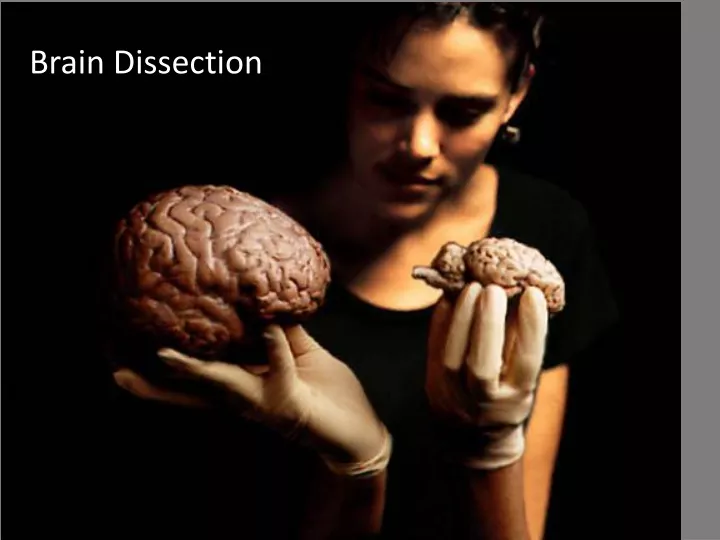
Brain Dissection. Orientation: Terms (different than humans). Dorsal. Cranial. Caudal. Ventral. External Observations. Part 1. Unless the directions specifically tell you to do so!. Preliminary observations. Measure the brain from the cranial to caudal end.

E N D
Orientation: Terms (different than humans) Dorsal Cranial Caudal Ventral
External Observations Part 1
Preliminary observations Measure the brain from the cranial to caudal end. Rinse sheep brain and pat dry. Weigh the brain and calculate size of the donor animal. (Sheep brain is ~ 0.5% of body weight; human brain is ~2% body weight) Record your observations.
The Meninges “Mater” • Examine the dura mater*, the toughest layer of the meninges covering and protecting the brain. Note how the dura mater wraps around the structures of the brain • The meningeal coverings are named descriptively: • Mater = mother • Dura = tough • Arachnoid = spider • Pia = gentle • The arachnoid mater, another layer of the meninges, is an ultra-thin spongy layer that may appear as a delicate “webby” material spanning the fissures or holding the dura mater to the brain. • The pia mater, another layer of the meninges, is the thin membrane that adheres tightly to the surface of the brain, dipping into each sulcus. If you pull away a small amount of the pia mater, you can see the depth of the sulcus.
Optic chiasma Pituitary gland Olfactory bulb This image shows the ventral surface of the sheep's brain; some of dura mater has been removed. Thepituitary gland and the optic chiasma (part of the cranial nerves) are still intact. (A = pituitary gland, B = optic chiasma, C = olfactory bulb)
Using a ruler approximate the sizes of the lobes • You can approximate their volume by measuring length, width and height. Parietal Lobe Occipital Lobe Frontal Lobe Temporal Lobe
Place the brain dorsal side up in the dissecting pan. Using scissors, gently cutthrough the dura mater between the cerebellum and the temporal lobe with the scissors pointing down toward the spinal cord. • Once the dura mater is free you should be able to carefully peel back the rest of the dura mater from the dorsal and lateral surface. Leave it in place on the ventral surface (SEE NEXT SLIDE FOR IMAGE). • You can now examine the dorsal and lateral structures of the brain. • The largest structure in the dorsal view is the cerebral cortex, the outer layer of the cerebrum. The cortex is folded upon itself, with a number of gyri (hills; singular = gyrus) and sulci (valleys; singular = sulcus).
Locate the four lobes of the cerebral cortex: • Frontal • Parietal • Temporal • Occipital • Locate the dividing lines between these lobes: the longitudinalfissureseparates the 2 hemispheres of the cerebral cortex, the centralsulcusseparates frontal lobe from parietal lobe; the lateralsulcusseparates parietal and temporal lobes (not shown in this photo). • Take a picture and label it. Sheep brain dorsal side; dura mater removed A – longitudinal fissure B - central sulcus C - frontal lobe D - parietal lobe E - occipital lobe F - temporal lobe G – cerebellum H – vermis of cerebellum I - medulla
Anterior to the central sulcus is the primary motor cortex. Posterior to the central sulcus is the primary sensory cortex. These correspond to the areas in the human brain. • Identify the cerebellum(little brain). In the human brain, this structure is divided longitudinally, whereas you will notice that in the sheep it is not. The fissures are also oriented differently in the sheep brain.
This lateral view shows the parietal lobe and cerebellum. If you were not careful when you removed the dura mater you may have accidentally pulled the entire cerebellum away from the brain. Note the transverse fissure, which separates the cerebrum from the cerebellum. The convolutions of the brain are also visible as bumps (gyri) and grooves (sulci).
Gently separate the 2 lobes of the cerebral cortex. DO NOT CUT! • Answer 4 in part 1 by probing the longitudinal fissure between the two hemispheres be careful to not pierce the soft tissue. • Place your finger on the probe where the top meets the brain, withdrawal the probe and measure the distance between the end of the probe and the location you marked on the probe with your finger
Parietal Occipital Temporal Cerebellum Frontal lobe Medulla Olfactory bulb & tract Optic chiasma Lateral View Sheep brain, lateral view; most of dura mater has been removed
You can also push the cerebellum down to show the pineal body from the side. It appears as a small, oval, slightly pink protrusion in the midline. The pineal body secretes melatonin, a hormone important in establishing day/night cycles, sleep induction and seasonal behavior (e.g., hibernation).
Ventral Structures Part 2
Sheep brain, ventral side, dura mater partially removed • Place the brain ventral side up in the dissecting pan. Remove all dura mater except for a small rectangle that leaves the pituitary and optic chiasma intact. • Take a picture of the ventral view and… • Use the following slides to locate the: optic chiasm, a cranial nerve, olfactory bulbs, pituitary gland, medulla, pons and spinal cord.
Frontal Lobe • Olfactory bulbs • Temporal Lobe • Optic Chiasm • 10. Pons • 12. Medulla Oblongata • 15. Optic Nerve (a cranial Nerve) • 16. Oculomotor Nerve (a cranial nerve) Not shown: pituitary
Look for the club-like olfactory bulbson the inferior surface of the frontal lobes of the cerebral hemispheres. • Measure the length of the olfactory bulbs and record.
Olfactory Bulbs Sheep Human
This picture, from the visible human project, shows the pathway of optic nerve from the back of the eye through the optic chiasm. • The optic nerve (II) carries sensory input from the retina of the eye to the brain. The X-shaped structure is where the fibers of each optic nerve cross over to the opposite side of the brain. • The optic tracts continue from the optic chiasm toward the occipital lobe – the primary visual center.
Pituitary Gland The pituitary gland, which produces important hormones, is a sac-like area that attaches to the brain between the pons and the optic chiasm. The pituitary gland will likely pull off if you remove the dura mater so be careful.
At the caudal border of the cerebral peduncles you will find the pons, followed caudal by the medullaoblongata. Finally, the spinal cord extends from the posterior portion of the medulla. The medulla ends (roughly) at the edge of the slightly rounded area, and at the beginning of the spinal canal (identified in the dorsal view). Pons Medulla Spinal cord
Cranial Nerves Part 3
Gently cutaway any remaining dura mater, being careful to preserve as many of the cranial nerves as possible. And the pituitary gland if possible The nerves will appear as white threads attached to the ventral surface of the brain. On this image, the dura matter has been completely removed, you can still see the optic chiasma but the pituitary gland is missing. The infundibulum (pituitary stalk) is now visible in the center. Careful dissection also reveals two other large nerves: the occulomotor nerves.
Identify as many of the cranial nerves as possible on your specimen and record your observations: • Olfactory bulb and tract • Optic chiasma / Optic nerve (II) • Trigeminal nerves (V) – involved in chewing and sensations of the head & face; this is a very large nerve tract on the sheep brain • Facial nerves (VII) - large nerves involved in taste sensation, gland function (salivary and lacrimal glands), and facial expressions • Glosso-pharyngeal nerve (IX) – swallowing, saliva production, taste buds; produces gag reflex • Vagus nerves (X) – serve many organs of the head, thorax & abdominal cavity (may be broken in your specimen)
Internal Structures Part 4
Turn the brain over so the dorsal side is up to make a sagittal cut. You are going to cut the brain in half along the line of the longitudinal fissure. • Bisect the brain along the longitudinal fissure, separating right and left hemispheres. You will be cutting through the corpus callosum. Hold your scalpel like a pencil and try to cut with one smooth slice, using enough pressure to cut with one smooth pass of the blade. If you do not get identical halves, ask for help to clean up the cut.
Before taking a picture of the bisected brain, use a probe to pull the choroid plexus out of the third ventricle (Sometimes it can be found in the lateral ventricle as well. • It will be dark pink in color. • Take a picture of the bisected brain
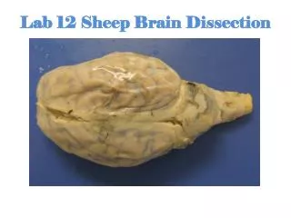
The image below shows a cleanly separated brain with the major internal structures visible.
Diencephalon • You can also see the thalamus, and the hypothalamus. The thalamus integrates information and relays it to appropriate regions for processing in the cerebrum. • The hypothalamus (“hypo” means below, so below the thalamus) in involved in many functions from biological timekeeping to most homeostatic regulation. The hypothalamus also controls the pituitary gland which is attached at its base. • The hypothalamus does not appear as a distinct structure, but is a general area bounded by other structures (thalamus, optic chiasm, and mammillary bodies).
Identify the cerebellumposterior to the fourth ventricle and notice the internal treelike arrangement of its white matter called the arbor vitae.
Coronal Section. Part 5
On the other half of your brain, make a Coronal (Frontal) section. Locate and label the cerebral cortex, grey mater, white mater, corpus collosum, and thalamus.
Clean up !! • Wrap your sheep brain in paper towels and dispose in the special bag as instructed by your teacher. • Clean all tools and your dissecting pan with warm soapy water. • Dry all equipment and put tools away. • WIPE THE KEYBOARD OF YOUR LAPTOP WITH DISPOSABLE WIPES BEFORE PUTTING IT AWAY!
This exercise not finished. Test yourself on the parts of the brain. Brain Parts Quiz: http://www.wiley.com/college/apcentral/anatomydrill/t14/at1401_1.htm Brain Function Quiz: http://www.wiley.com/college/apcentral/anatomydrill/t14/at1422_1.htm
Thank you for your interest in sheep and their amazing brains!