Download
1 / 40
400 likes | 573 Views
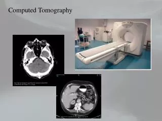
Computed Tomography RAD309. Data Acquisition. Data Acquisition. Data acquisition represents the first step in process of image production X-ray tube & detectors collect information systematically Collect large number of x ray transmissions around the patient. Data Collection Basics. Patient.

E N D
Computed TomographyRAD309 Data Acquisition
Data Acquisition • Data acquisition represents the first step in process of image production • X-ray tube & detectors collect information systematically • Collect large number of x ray transmissions around the patient
Data Collection Basics Patient • X-ray source & detector must be in & stay in alignment • Beam moves (scans) around patient • many transmission measurements taken X-Ray beams
Data Collection Basics • Pre-patient beam • collimated to pass only through slice of interest • shaped by special filter for uniformity
Data Collection Basics (cont) • Beam attenuated by patient • Transmitted photons detected by scanner • Detected photon intensity converted to electrical signal (analog) • Electrical signal converted to digital value • A to D converter • Digital value sent to reconstruction computer
CT “Ray” • That part of beam falling onto a single detector Ray
Each CT Ray • attenuated by patient • projected onto one detector • detector produces electrical signal • produces single data sample
CT Projection -or- View • # of simultaneously collected rays
Acquisition Geometries • Pencil Beam • Fan Beam • Spiral
DA Geometries • Parallel beam, translate rotate motion • Fan beam, translate rotate motion • Fan beam, complete rotation tube/detector • Fan beam, complete rotation of tube around stationary ring of detectors • Special: high speed CT, stationary/stationary, multiple targets tube • Spiral, rotate/translate • Multiple detector rows
Interconnect Wiring Tube Slip Rings Detector Spiral Geometry • X-ray tube rotates continuously around patient • Patient continuously transported through gantry • No physical wiring between gantry & x-ray tube • Requires “Slip Ring” technology
X Ray System • Initially used low energy gamma rays • Problem: low radiation intensity rate, large source size, low source strength, high cost • Use of X ray tubes • Benefit: high radiation intensity, high contrast ct scanning • Problem: heterogeneous beam , does not obay Lamber-Beer Exponential Law
Radioactive Source instead of an X-Ray Tube? • High intensity required • X-ray tubes produce higher intensities than sources • Single energy spectrum desired • Produced by radioactive source • X-ray tubes produce spectrum of energies
CT Beam Filtration Shapes beam to appear monochromatic and satisfy reconstruction process • Hardens beam • Removes greater fraction of low-energy photons than high energy photons • reduces patient exposure • Shapes energy distribution to produce uniform intensity & beam cross section Patient Filter
Patient Protection • Pre-collimators • between tube & patient Tube • Post-collimators • between patient & detector Detector
Tube Detector Pre-Collimation • Constrains size of beam • Reduces amount of scatter produced • Designed to minimize beam divergence • Often consists of several stages or sets of jaws Pre-collimator
Tube Detector Post-Collimation • Helps define slice (beam) thickness • Reduces scatter radiation reaching detector • Improves image quality Post-collimator
Detectors • Capture radiation from patient • Converts to electrical signal • Then they are converted to binary coded information
CT Detector Characteristics • Efficiency • Response time • Dynamic range • Reproducibility and Stability
1. Efficiency • Ability to capture, absorb & convert x-ray photons to electrical signals
Efficiency Components a. Capture efficiency • Efficiency of detector to obtain transmitted photons from patient • Size of detector area, distance between 2 detectors b. Absorption efficiency • no. of photons absorbed • Z , density, size, thickness of detector c. Conversion efficiency • fraction of absorbed energy which produce signal
Overall Detector Efficiency capture efficiencyXabsorption efficiencyXconversion efficiency
Absorption Efficiency • Depends upon detector’s • atomic # • density • size • thickness • Depends on beam spectrum
2. Response Time • “Speed with which detector can detect an x ray event and recover to detect the next one” • Minimum time after detection of 1st event when detector can detect 2nd event • If time between events shorter than response time, second event may not be detected • Shorter response time better
3. Dynamic Range • Ability to faithfully detect large range of intensities • “Ratio of largest signal to be measured to the precision of the smallest signal to be discriminated” • Typical dynamic range: 1,000,000:1 • much better than film
4. Stability • “Steadiness” of detector system • Consistency of detector signal over time • The less stable, the more frequently calibration required
Detector Types 2 principles: • Convert x-ray into light ---electrical signal • Scintillation detector • Convert x-ray directly into electrical signal • Gas ionization detector
Scintillation Detectors • Crystal couple to photomultiplier tube • X ray falls on crystal ---light flashes (glow) • Light directed to PM • Light hits Photocathode in PM and releases electrons
Light X-Rays Scintillation • X-ray energy converted to light • Light converted to electrical signal Photomultiplier Tube Electrical Signal Scintillation Crystal
Photomultiplier Tubes • Light incident on Photocathode of PM tube • Photocathode releases electrons + - Light X-Rays Scintillation Crystal Photocathode PM Tube Dynodes
Gas Ionization Detector • Series of individual chambers separated by tungsten plates • X ray falls on each chamber– (+/- ions) • + ions move to – plate, - ions to + plate • The migration produces electrical signal
Electrical Signal X-Rays Gas Ionization • X-rays converted directly to electrical signal Filled with Air Ionization Chamber + - - +
CT Ionization Detectors • Many detectors (chambers) used • adjacent walls shared between chambers • Techniques to increase efficiency • Increase chamber thickness • x-rays encounter longer path length • Pressurize air (xenon) • more gas molecules encountered per unit path length thickness X-Rays
Detector Array • Slice by Slice – one arc of detector array • Volume – one arc of detector array, acquires volume of tissue then separated by computed to slice by slice
DAS • Detector electronics • Location: between detector and computer • Role of translator • Measure transmitted radiation beam • Encodes measurement to binary data • Transmits binary data to computer
Components of DAS • Amplifier • Log Amplifier • Analog to Digital Converter (digital data) • Digital Transmission to computer
Log Amplification • Transmission measurement data must be changed into attenuation and thickness data • Attenuation = log of transmission x thickness
Detector Electronics From Detector Increases signal strength for later processing Amplifier Compresses dynamic range; Converts transmission intensity into attenuation data Logarithmic Amplifier Analog to Digital Converter To Computer
DA and Sampling • Radiation falling on detector • Each samples the beam intensity on it • Not enough samples = artifacts appear • To increase number of measurement/samples available for reconstruction and improve image quality
Improving Quality & Detection • Geometry • Smaller detectors • Closer packed detectors • Smaller patient-detector distance • Thinner slices • less patient variation over slice thickness distance