Download

1 / 52
520 likes | 718 Views
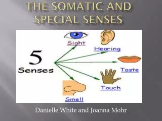
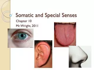
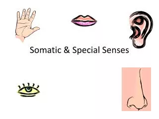
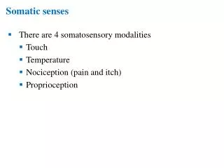
Somatic and Special Senses. Somatic Senses Touch Pressure Temperature Pain. Special Senses Smell Taste Hearing Equilibrium vision. Categories. Types of Receptors. Chemoreceptors – stimulated by changes in the chemical concentration of substances Pain receptors - … by tissue damage

E N D
Somatic Senses Touch Pressure Temperature Pain Special Senses Smell Taste Hearing Equilibrium vision Categories
Types of Receptors • Chemoreceptors – stimulated by changes in the chemical concentration of substances • Pain receptors - … by tissue damage • Thermoreceptors - … by changes in temp. • Mechanoreceptors - … by changes in pressure or movement • Photoreceptors - … by light energy
Sensation • A feeling that occurs when the brain interprets sensory impulses • Depends on which region of the brain receives the impulse • Sensory adaptation – ability to ignore unimportant stimuli
Somatic Senses • Senses of touch and pressure • Free nerve endings (touch and pressure) • Meissner’s corpuscles (light touch) • Abundant in hairless portions of body • Pacinian corpuscles (heavy pressure)
Temperature senses • Warm receptors • Sensitive to 77°F and above; become unresponsive at temperatures above 113°F • At or above 113°F stimulates pain receptors, producing a burning sensation • Cold receptors • Sensitive 50°F to 68°F • Below 50°F stimulate pain receptors, producing a freezing sensation • Sense of pain • Widely distributed, except for brain • Protect the body b/c tissue damage stimulates them • Pain is perceived as unpleasant and it signals person to act to remove the simulation • Pain receptors adapt poorly
Visceral pain • Localized damage to intestinal organs may not elicit pain sensations • More widespread stimulation, as when intestinal walls are stretched, a strong pain sensation may follow • May feel as though it is coming from some part of the body other than the part being stimulated, a phenomenon known as referred pain
Regulation of Pain Impulses • Awareness arises when impulses reach the thalamus • Cerebral cortex determines pain intensity, locates pain’s source, and mediates emotional and motor responses to the pain • Graymatter in midbrain, pons, and medulla oblongata regulate movement of pain impulses from the spinal cord • Inhibiting substances released (enkephalins, serotonin, endorphins)
Olfactory organs contain the olfactory receptors and are yellowish-brown masses of epithelium about the size of postage stamps that cover the upper pars of the nasal cavity Sense of Smell
Olfactory receptors are bipolar neurons surrounded by columnar epithelial cells • There are 400 types of olfactory receptor proteins • Odorant molecules enter the nasal cavity as gases, dissolve in watery fluids, and then bind to the receptors in different patterns • Stimulated olfactory receptors cells send nerve impulses along their axons which synapse with neurons located in enlargements called olfactory bulbs
Within the olfactory bulbs, the impulses travel along the olfactory tracts to the limbic system within the temporal lobe and base of the frontal lobes in the brain • Sense of smell adapts rapidly but adaptations to one scent will not diminish sensitivity to new odors
Humans have about 12 million olfactory receptor cells • Bloodhounds have 4 billion olfactory receptor cells
Taste buds are special organs of taste Our 10,000 taste buds are located primarily on surface of the tongue and are associated with tiny elevations called papillae About 1,000 taste buds are scattered in the roof of the mouth and walls of the throat Sense of Taste
Taste receptors • Each taste bud includes a group of modified epithelial cells, the gustatory cells, which function as receptors cells • Each taste bud has 50-150 receptor cells • Entire structure is spherical with an opening called the taste pore and projections called taste hairs which are the sensitive parts • Nerve fibers wrap around the taste cells • Stimulation of taste cell triggers an impulse on a nearby nerve fiber and the impulse travels into the brain
The truth about cats and dogs… • They can be satisfied with less varied diets than humans • Cats have 473 taste buds • Dogs have 1,700 taste buds
Tasting • Chemical must be dissolved in watery fluid provided by salivary glands • Food molecules bind to specific receptors embedded in and projecting from taste hairs on the taste cells • Pattern of receptor types generate sensory impulses on nearby nerve fibers in interpreted as a particular taste sensation
Taste sensations Primary taste sensations Sweet Sour Salty Bitter Others recognized Alkaline Metallic Umami
Flavor • Results from one or a combination of the primary sensations • Experiencing flavors involves tasting, smelling, and feeling the texture and temperature of foods • Some foods, such as chili peppers and ginger, trigger heat receptors • Taste sensation undergoes adaptation rapidly • Moving bits of food over the surface of the tongue to stimulate different receptors at different moments keeps us from losing taste
Outer ear Auricle (funnel-like structure) External auditory meatus (ear canal) tympanic membrane (ear drum) Sense of Hearing
Middle ear • Air-filled space in the temporal bone • Contains 3 small bones called auditory ossicles: • Malleus • Incus • Stapes • Malleus attaches to eardrum and when the eardrum vibrates, the malleus vibrates in unison • The malleus causes the incus to vibrate, and the incus passes the movement on to the stapes • Vibration of the stapes at the oval window moves a fluid within the inner ear, which stimulates the hearing receptors
Auditory ossicles also help increase (amplify) the force of vibrations • The pressure that the stapes applies on the oval window is many times greater than the pressure that sound waves exert on the eardrum
Auditory tube (eustachian tube) • Connects each middle ear to the throat • Conducts air between the tympanic cavity and the outside of the body by way of the throat • Auditory tube helps maintain equal air pressure on both sides of the eardrum, which is necessary for normal hearing • Ex. Rapid changes in altitude may push eardrum inward as air pressure on the outside of the eardrum incerases
Steps in the generation of sensory impulses from the ear • Sound waves enter external acoustic meatus • Waves of changing pressure cause eardrum to reproduce vibrations coming from sound wave source • Auditory ossicles amplify and transmit vibrations to end of stapes • Movement of stapes at oval window transmits vibrations to perilymph in scala vestibuli • Vibrations pass through vestibular membrane and enter endolymph of cochlear duct
Different frequencies of vibration in endolymph stimulate different sets of receptor cells • As a receptor cell depolarizes, its membrane becomes more permeable to calcium ions • Inward diffusion of calcium ions causes vesicles at the base of the receptor cell to release neurotransmitter • Neurotransmitter stimulates ends of nearby sensory neurons • Sensory impulses are triggered on fibers of the cochlear branch of vestibulocochlear nerve • Auditory cortex of temporal lobe interprets sensory impulses
Factors of hearing loss • Conductive deafness • Interference with the transmission of vibrations to the inner ear • May be due to plugging of the external acoustic meatus or to changes in the eardrum or auditory ossicles • Eardrum may harden as a result of disease and thus be less responsive to sound waves • Disease or injury may tear or perforate the eardrum • Sensorineural deafness • Damage to the cochlea, auditory nerve, or auditory nerve pathways • Causes by loud sounds, tumors in the CNS, brain damage as a result of vascular accidents, or use of certain drugs
Sense of Equilibrium • Static equilibrium • Sense the position of the head, maintaining stability and posture when the head and body are still • Organs located with the vestibule, a bony chamber between the semicircular canals and the cochlea • A tiny structure called a macula contains numerous hair cells which serve as sensory receptors
When the head is upright, the hairs of the hair cells project upward into a mass of gelatinous material, which has grains of calcium carbonate embedded in it • Head bending tilt the gelatinous mass and they sag in response to gravity • Hairs within the mass bends and they signal nerve fibers • Nerve impulses travel into the CNS and informs brain of head’s new position • Brain responds by sending motor impulses to skeletal muscles, which contract or relax to maintain balance
Dynamic equilibrium • Aid in maintaining balance when the head and body suddenly move or rotate • Organs are the three semicircular canals • Each responds to a different anatomical plane • Each canal ends in a swelling called an ampulla which contains the sensory organs crista ampullaris • Each of the crista ampullaris contains a number of sensory hairs cells which extend upward in a gelatinous mass • Rapid turns of the head or body simulate these hair cells
Other sensory structures that aid in maintaining equilibrium • Mechanoreceptors associated with joins • Eyes detect changes in posture
Motion sickness • Nausea, vomiting, and headache • Arise from senses that don’t make sense • Eyes of person reading in a moving car, for example, signal the brain that the person is stationary, because the print doesn’t move • Skin and inner ear, however, detects movement • To lessen the misery, focus on the horizon or an object in the distance ahead or take dramamine
Sense of Sight • Visual accessory organs • Eyelid • Skin • Thinnest skin in the body • Covers the lid’s outer surface and fuses with its inner lining near the margin of the lid • Muscle • Eyelids moved by orbicularis oculi and levator palpebrae superioris • Conjuctivia • Mucous membrane that lines the inner surfaces of the eyelids and fols back to cover the anterior sruface of the eyeball, except for its central portion (cornea)
Cornea • Lens • Fornix • Marginal conjuctiva • Lacrimal glands • Tarsus, a fibrous connective tissue • Lacrimal glands • Secretes tears • Moistens and lubricates the surface of the eye and the lining of the lids • Contain lysozyme that is an anti-bacterial agent, reducing the risk of eye infections
Extrinsic muscles • Arise from the bones of the orbit and insert by broad tendons on the eye’s tough outer surface • Six muscles move the eye in various directions
Structure of the eye • Hollow, spherical structure about 2.5 cm in diameter • Wall has 3 distinct layers • Outer fibrous layer • Middle vascular layer • Inner nervous layer • Spaces within the eye are filled with fluids that support its wall and internal parts and help maintain its shape
Outer layer • Outermost layer (1/6 of layer) is transparent cornea, which is the window of the eye and helps focus entering light rays • Composed largely of connective tissue • Very few cells and no blood vessels • Along its circumference, the cornea is continuous with the sclera, the white portion of the eye (5/6 of layer) • Opaque due to many large collagenous and elastic fibers • Sclera protects the eye and is an attachment for the extrinsic muscles • The optic nerve and blood vessels pierce the sclera in the back of the eye
Most common cause of blindness is loss of transparency of the cornea • A corneal transplant can replace the central 2/3 of the defective cornea with a similar-sized portion of cornea from a donor eye
Middle layer • Choroid coat • Posterior 5/6 of the globe of the eye • Contains blood vessels which nourish surrounding tissue • Contains melanocytes • Melanin absorbs excess light and keeps the inside of the eye dark • Ciliary body • Extends forward from the choroid coat and forms and internal ring around the front of the eye • Contains ciliary muscles • Lens • Transparent and held in place by suspensory ligaments • Adjusts shape to facilitate focusing, called accommodation
Cataracts • Lens or its capsule slowly becomes cloudy and opaque • Without treatment, they cause blindness • Causes by breakdown of protein within lens after age 45
Iris • Thin diaphragm composed mostly of connective tissue and smooth muscle fibers • Colored portion of the eye • Lies between the cornea and the lens • Aqueous humor • Watery fluid secreted by the ciliary body • Circulates through the pupil (circular opening in the center of the iris) and into the anterior chamber • helps nourish cornea and lens and aids in maintaining the shape of the front of the eye
Smooth muscle fibers of iris are organized into two groups, a circular set and a radial set • When circular set contracts, pupil gets smaller and less light enters • When the radial set contracts, the pupil’s diameter increases and more light enters
Glaucoma Disorder in which rate of aqueous humor formation exceeds rate of its removal Fluid pressure squeezes shut blood vessels that supply the receptor cells of the retina Cells that are robbed of nutrients and oxygen may die and permanent blindness can result Treated with drugs, laser therapy, or surgery
Inner layer • Retina • Contains the visual receptor cells (photoreceptors) • Continuous with the optic nerve and extends forward as the inner lining of the eyeball • Central region is a yellowish spot called the maculla lutea • Depression in its center is called the fovea centralis which is where the sharpest vision occurs • Optic disc is just medial to the fovea centralis and contains a central artery and vein which are continuous with the capillary networks • Space bounded by the lens, ciliary body, and retina is the largest compartment and is filled with a transparent, jellylike fluid called vitreous humor • Vitreous humor helps eye maintain shape
Light refraction • When a person sees something, either the object is giving off light or light waves are reflected from it • These light waves enter the eye and an image of the object is focused on the retina • Focusing bends the light waves, a phenomenon called refraction • Convex surface of the cornea refracts light waves from outside objects • Convex surface of lens then refracts the light again • If eye shape is normal, light waves focus sharply on the retina • Image that forms is upside down and reversed from left to right • Visual cortex interprets the image in its proper position
Visual receptors • Modified neurons • Rods • Have long, thin projections at their ends • Provide black and white vision • Provide general outline of objects • 100 times more sensitive to light than cones • Humans have 125 million of them • Cones • Short, blunt projections • Provide color vision • Provide sharp images • Humans have 7 million of them • Stimulated only when light reaches them