Download
1 / 78
790 likes | 951 Views
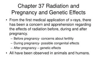
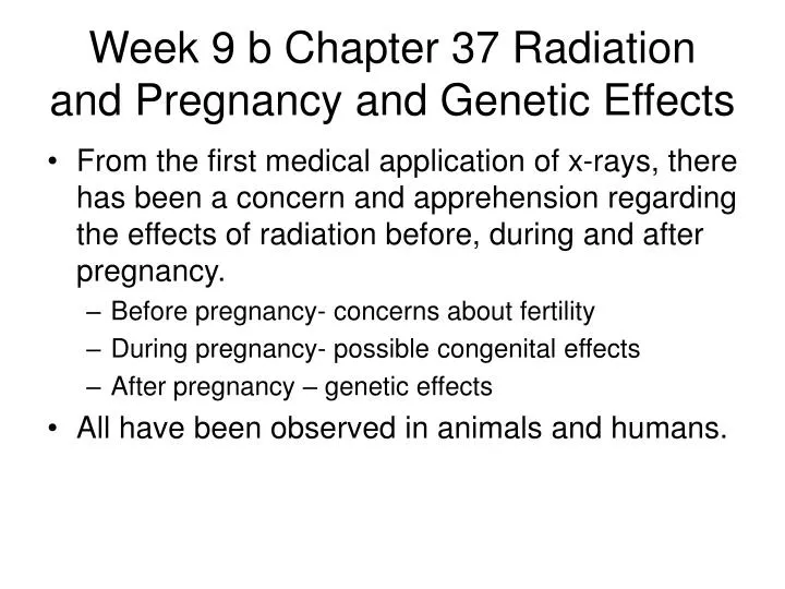
Week 9 b Chapter 37 Radiation and Pregnancy and Genetic Effects. From the first medical application of x-rays, there has been a concern and apprehension regarding the effects of radiation before, during and after pregnancy. Before pregnancy- concerns about fertility

E N D
Week 9 b Chapter 37 Radiation and Pregnancy and Genetic Effects • From the first medical application of x-rays, there has been a concern and apprehension regarding the effects of radiation before, during and after pregnancy. • Before pregnancy- concerns about fertility • During pregnancy- possible congenital effects • After pregnancy – genetic effects • All have been observed in animals and humans.
Effects on Fertility • The early effects of high level radiation exposure on fertility is known in both men and women. • Doses as low as 10 rad may delay or suppress menstruation in women and reduce the number of spermatozoa. • Doses of 200 rad can produce temporary infertility. • Dose of 500 rad will produce sterility.
Effects on Fertility • The short term effects are documented and known to be dose related. • The effects of low-dose, long term irradiation on fertility is less well defined. • Animal results are lacking. Those that exist demonstrate no effect in exposure of 100 rad per year. • A study of 150,000 rad techs found no effect on fertility over a 12 year sampling period. • Low-dose, chronic irradiation does not impair fertility.
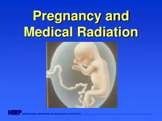
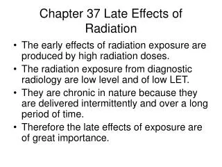
Irradiation in Utero • Irradiation in utero concerns two types of exposures. • Those to the radiation worker • Those to the patient • There is substantial data about animal exposure to relatively high doses of radiation delivered during the various periods of gestation.
Irradiation in Utero • The embryo is a rapidly developing cell system so it is very sensitive to radiation. • With age, the radiosensitivity decreases and the pattern continues into adulthood. • The effects of exposure in utero are time and dose related.
LD50/60 of mice • After maturity, radiosensitivity increases with age. • It begins to decrease with age at the end of child bearing age. • The study shows mice age in weeks and human age in years.
Exposure During the 1st Trimester • All observations point to the first trimester of pregnancy as the most radiosensitive period. • With in the first two weeks after fertilization to most pronounced effect of a high radiation dose is fetal death which is manifested as a spontaneous abortion. • Observed in radiation therapy patients but only after very high exposures. • The 1st 2 weeks of pregnancy may be of least concern because the response is all or nothing.
Exposure During the 1st Trimester • From animal data, it would appear that this response is very rare. The best estimate is a 10 rad exposure during the first two weeks would induce 0.1% rate of spontaneous abortion. This is added to the 25% to 50% normal incidence of spontaneous abortion. • Fortunately there will either be a spontaneous abortion or the pregnancy will carry to full term with no effect.
Exposure During the 1st Trimester • During the 2nd to 10th weeks two effects may occur. • Early in this period, skeletal and organ abnormalities can be induced. • As organs continue to develop, central nervous system abnormalities may develop. • If the abnormalities are severe enough, the effect will be fetal death. • After a dose of 200 rad, nearly 100% of the fetuses suffered significant abnormalities. • In 80%,it was sufficient to cause death.
Exposure During the 1st Trimester • Such effects after diagnostic levels of exposure are essentially undetectable after radiation doses of less than 10 rad. • A dose of 10 rad during organogenesis is expected to induce congenital abnormalities by 1% above natural occurrence. • To complicate this there is a 5% incidence of naturally occurring congenital abnormalities in the unexposed population.
Exposure During the 1st Trimester • In utero irradiation has been associated with childhood malignancy. Oxford University has studies every childhood malignancy in England, Scotland and Wales since 1946. • They found that childhood leukemia was of particular importance. The relative risk was 1.5 or an increase of 50% over the non-irradiated rate. The number is very small.
Time of X-ray Exam First Trimester Second Trimester Third Trimester Total Relative risk 8.3 1.5 1.4 1.5 Relative risk of Childhood Leukemia after Irradiation by Trimester
More Effects of in Utero Exposure • Observed in Atomic Bomb survivors has been an increase in mental retardation. This involved very high exposure rates • It is known that radiation does retard growth. Irradiation particularly during organogenesis has been associated with microcephaly (small head) and retardation. • Human data from atom bomb survivors and residents of the Marshall Islands exposed to fall out from atom bomb tests demonstrated impaired growth in children.
Genetic Effects • We have no data suggesting that radiation induced genetic effects in humans. There has been no observed radiation induced genetic effects in the atomic bomb survivor or Marshall Islanders. They are now in their third generation. • We must therefore rely on work done on fruit flies and mice.
H.J. Muller work with Fruit Flies • Nobel prize winning geneticist H.J. Muller irradiated mature fruit flies and then measured the frequency of lethal mutations. • He used thousands of rad but the response was linear non-threshold.
H.J. Muller work with Fruit Flies • It was the results of his work that in 1932 the National Council on Radiation Protection (NCRP) lowered the recommended dose limits for workers and acknowledged the existence of the linear non-threshold radiation effects. • Since that time all radiation protection guides have assumed a linear non-threshold response.
Conclusions Regarding Radiation Genetics • Radiation-induced mutations are usually harmful. • Any dose of radiation, however small, to a germ cell results in some genetic risk. • The frequency of radiation induced mutations is directly proportional to dose, so that a linear extrapolation of data from high exposures proves a valid estimate for low dose effects.
Conclusions Regarding Radiation Genetics • The effect depends upon protraction and fractionation. • For most of the preproductive life, the women is less sensitive to genetic effects of radiation than the man. • Most radiation-induced effects are recessive. This requires the mutant gene to be present in both the male & female to produce the trait. Such mutations may not be expressed for many generations. • The frequency of radiation induced genetic mutations is extremely low. It is approximately 10-17 mutations/rad/gene.
Conclusion for Diagnostic Exposures • The incidence of radiation-induced genetic mutations from exposure levels in diagnostic radiology is zero. • For nearly all diagnostic exposures, no action is required. • Should a high exposure in excess of 10 rad, some protective action may be required.
Female Precautions • The prefertilized egg, in its various stages exhibits a constant sensitivity to radiation. • It has demonstrated the ability to repair genetic damage. • If repairs occur, it is rapid and therefore a delay in procreation of only a few days may be prudent.
Male precautions • In the male, it might be prudent to refrain from procreation for a period of 60 days to allow cells that were in a resistant stage of development at the time of exposure to mature to functioning spermatids
Summary • The effects of low-dose, long term irradiation in utero can include • Prenatal death • Congenital abnormalities • Malignancies • Impairment of growth • Mental retardation • Genetic Effect
Summary • These abnormalities are based upon exposure of more than 100 rad in humans and greater than 10 rad in animal experiments. • There is no evidence that diagnostic levels of radiation exposure currently experienced occupationally or medically are responsible for any such effects on fetal growth or development.
Chapter 38 Health Physics • The term health physics was coined during the Manhattan Project as they developed the atomic bomb. • A group of physicists and physicians were responsible for the radiation safety of personnel involved in the production of the atomic bombs. • The health physicist is concerned with: • Research • Teaching • Operational aspects of radiation safety.
Health Physics • Health physics is concerned with providing occupational radiation protection and minimizing radiation dose to the public. • There are three cardinal principles developed for nuclear activities that have equally useful applications in diagnostic radiology. • By observing these principles, radiation exposure can be minimized.
Cardinal Principles of Radiation Protection • Keep the time of exposure to radiation as short as possible. • Maintain a large distance as possible between the source of radiation and the exposed person. • Insert shielding material between the radiation source and the exposed person.
Minimize Time • Exposure = Exposure rate x exposure time • During radiography the exposure time is reduced to reduce motion blur. • During fluoroscopy exposure time is reduced to reduce patient and personnel exposure. Radiologists are trained to switch the exposure on and off rather than continuous on to lower exposure. Pulsed progressive fluoroscopy reduces patient exposure by a factor of 0.1 or less • Fluoroscopy machine have a 5 minute reset timer.
Maximize Distance • As the distance from the source of radiation increases, the radiation exposure rapidly decreases. inverse square law • I1 D22 • ---- = ------- • I2 D12 • The source of radiation can be a point source like the tube and an extended area source like the patient. • If you are 5 times the source diameter from the sources, it can be treated as a point source.
Maximize Distance • During radiography the operator should be completely within a shielded control booth. • During stress radiography, the operator may need to manipulate the area being radiographed so they should be in a lead apron and lead gloves. • They also should be as far away from the primary beam as possible.
Maximize Distance • During normal radiography, only the patient should be in the room during exposures. • During fluoroscopy, the operator and technologist are in the room during exposure. • During fluoroscopy the technologist should be as far away from the patient as possible.
Maximize Distance • The exposure rate can be computed using isoexposure line to determine the safest place to stand during an exam.
Maximize Shielding • Positioning shielding between the radiation source and exposed persons greatly reduces the level of radiation exposure. • Shielding in radiography is generally lead or concrete. • The amount of shielding material needed can be estimated if we know the HVL Half Value Layer or Tenth Value Layer of the shielding material.
Maximize Shielding • The HVL is the amount of material needed to reduce intensity by 50%. • The TVL is the amount of absorber needed to reduce intensity to 10% of the original value • 1TVL = 3.3 HVL
Approximate HVL & TVL of Lead and Concrete at Various Tube Potentials
Dose Limits • A continuing effort of the health physicists is to describe and identify occupational dose limits. • For years a Maximum Permissible Dose (MPD) was specified for radiation workers. • The MPD was the dose of radiation that would be expected to produce no significant radiation effects.
Dose Limits • At radiation levels below the MPD, no response should occur. That’s the problem with MPD. • The response at or below the MPD is not zero because of the linear non-threshold radiation dose response. • The MPD has been replaced by Dose Limits (DL).
Dose Limits • The NCRP has assessed risks based upon the BEIR Committee and the National Safety Council. • State and federal government agencies routinely adopt the recommended dose limits as law. • They are developed for whole body and various organs and for various working conditions.
Radiation Dose Limits • Particular care must be taken to make certain that no radiation worker receives a radiation dose in excess of the DL. • The DL is specified for occupational exposures. • It must not be confused with medical x-ray exposure as a patient they receive. • Patient exposure is kelp as low as possible but there is no limit or DL for patients.
Radiation Dose Limits • The DL of 50,000 mrem per week was established in 1902. Through the years there has been a downward revision to the dose limits. • The early DL was based upon a known acute response level and presumed that a threshold dose existed. • The NCRP limits established in 1987 is 50 mSv per year (5000 mrem) Cumulative 10 mSv x age • The International Commission on Radiation Protection limits established in 1991 limits of 20 mSv per year (2000 mrem)
Radiation Dose Limits • Because the basis of the DL assumes a linear, nonthreshold response, all unnecessary radiation must be avoided. • Occupational exposure is defined as dose equivalent in units of millisevert (millirem). • DL are specified as Effective Dose (E) • The effective dose concept accounts for different types of radiation with varying relative biologic effectiveness. • Effective dose also considers the relative radiosensitivity of various tissue and organs.
Radiation Dose Limits • The effective dose concept is very important consideration when protective apparel such as lead aprons are used. • Wearing an apron effectively reduces the radiation dose to many tissue types and organs to nearly zero. • Therefore the effective dose is much less than the dose measured at collar level with a film badge.
Effective Dose • Effective dose (E) = radiation weighting factor (Wr) times Tissue weighting factor (Wt) time the Absorbed dose. • For medical radiation uses, the radiation weight factor is 1. For other type of radiation it is based upon the LET of the radiation. • The tissue weighting factor ranges from 0.20 for gonads (most radiosensitive) to 0.01 for skin (less radiosensitive).
Dose Limits for Tissues and Organs • The whole body DL of 50mSv/year is an effective dose which take into account the weighted average of various tissue types and organs. • Skin Some organs have a higher DL than the whole body DL. The DL for skin is 500 mSv/year. • Eyes The DL for eyes is 15mSv/year.
Public Exposure • Individuals in the general population are limited to 5mSv/year(500 mrem/year) if the exposure is infrequent. If the exposure is frequent as with a hospital employee who may visit radiology, the limit is 1 mSv/year (100 mrem/year). • The 1mSv/year DL is what physicist use to compute thickness of protective barriers.
Educational Considerations • Student under the age of 18 may not receive more than 1 mSv/year during their course of educational activities. • For this reason, student technologists under 18 may be engaged in x-ray imaging but their exposure is limited to 1mSv/year. • It is a general practice to not accept underage students for RT programs.
ALARA Considerations • ALARA stands for As Low As Reasonably Achievable and is the corner stone of radiation safety policies and procedures. • The 1991 ICRP recommendation of reducing annual exposures to 20mSv per year is under review by the NCRP.
DL’s, X-rays and Pregnancy • Two situations in diagnostic radiology require particular care and action. Both are associated with pregnancy. Their importance is obvious from both a physical and emotional stand point. • The severity of potential response to radiation in utero is both time and dose related.
X-rays during the first two weeks of pregnancy. • A grave misconception is that the most critical time for irradiation is during the first two weeks of pregnancy when it is unlikely that the mother knows that she is pregnant. If fact this time during pregnancy is the least hazardous. • The most likely biologic response to irradiation during the first two weeks is resorption of the embryo and therefore no pregnancy. • There is a concern about possibility of inducing congenital abnormalities during this time but it has not been demonstrated in animals or humans at any level of radiation exposure.