Download

1 / 37
390 likes | 793 Views
Large Intestine & Inferior Mesenteric Artery . Objectives. Discuss anatomical structure of large intestine. Enlist the characteristic features of large intestine. What are the different positions of the Appendix. Describe the blood supply of the large intestine.

E N D
Objectives • Discuss anatomical structure of large intestine. • Enlist the characteristic features of large intestine. • What are the different positions of the Appendix. • Describe the blood supply of the large intestine.
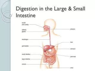
The large bowel may vary considerably in length in different subjects;the average is approximately 5 feet (1.5 m). • The large intestine is subdivided, for descriptive purposes, into: • Caecum with the Appendix vermiform • Colon ascending colon hepatic flexure transverse colon splenic flexure descending colon sigmoid colon • Rectum & Anal canal .
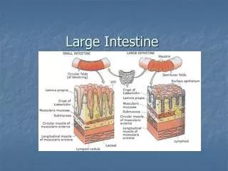
The general characteristics of most of the large intestine are: • Its large internal diameter compared to that of the small intestine; • theappendices epiploicae (omental appendices) are fat-filled peritoneal tags • The taeniae coli: three thickened bands of muscles • the haustra of colon are sacculations of the colon between the taeniae
No taeniae in the appendix or rectum. • The colon (but not the appendix, caecum or rectum), bears characteristic fat-filled peritoneal tags called appendices epiploicae scattered over its surface. • These are especially numerous in the sigmoid colon. • The transverse colon and sigmoid are completely peritonealized (the former being readily identified by its attachment to the greater omentum).
The ascending and descending colon have no mesocolonbut adhere directly to the posterior abdominal wall . • The caecum is usually completely peritonealized, • The appendix has its own mesocolon.
Features of large intestine: Taeniae Coli:Three thickened bands of muscles No taeniae in the appendix or rectum Haustra: Sacculations of the colon between the taeniae Omental Appendices: Small fatty projections of the omentum Caliber: The internal diameter is much bigger than small intestine
Cecum and Appendix Ileocecal Junction Taenia Coli Sacculations = Haustra
The cecumis that part of the large intestine that lies below the level of the junction of the ileum with the large intestine . It is a blind-ended pouch that is situated in the right iliac fossa. It is about 2.5 in. (6 cm) long and is completely covered with peritoneum. • The appendix is attached to the posteromedial wall of the cecum, just inferior to the end of the ileum
The appendix is suspended from the terminal ileum by the mesoappendix, which contains the appendicular vessels . • Its point of attachment to the cecum, the base of the appendix, is consistent with the highly visible free taenia leading directly to it. • But the location of the rest of the appendix varies considerably . • The appendix is at the junction of the lateral and middle one-thirds of a line from the anterior superior iliac spine to the umbilicus (McBurney's point).
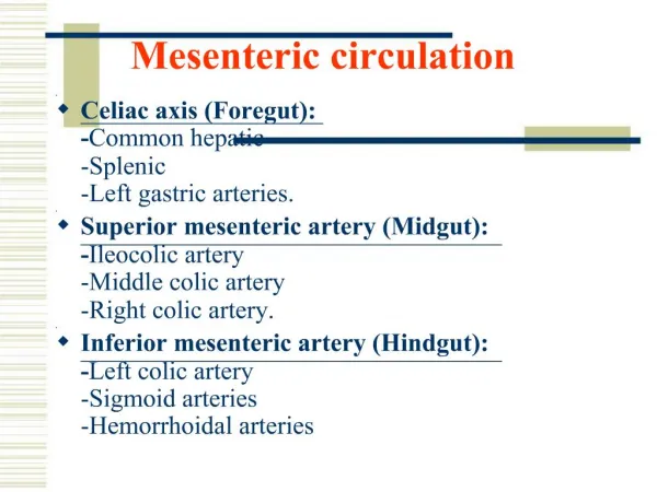
The inferior mesenteric artery and arises anterior to the body of vertebra L3. Its branches include the • left colic artery, • several sigmoid arteries, • superior rectal artery. • The veins drain into the inferior mesenteric vein