Download
1 / 36
390 likes | 707 Views
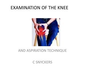
Knee Examination. Dr M.Kumar & Dr A. Osman HDR 23/10/2013. History:. Pain . 'popping' or 'snapping' sound may suggest rupture of a ligament. Swelling: rapid swelling (0-2 hours) e.g haemarthrosis which may, be due to ACL or PCL rupture, and patellar dislocation.

E N D
Knee Examination Dr M.Kumar & Dr A. Osman HDR 23/10/2013
History: • Pain. • 'popping' or 'snapping' sound may suggest rupture of a ligament. • Swelling: rapid swelling (0-2 hours) e.g haemarthrosis which may, be due to ACL or PCL rupture, and patellar dislocation. • Gradual swelling (6-24 hours) suggests an effusion, e.g meniscal injury. • Swelling over a 24-hour, with no history of trauma, ? septic arthritis ? inflammatory arthritis. • Locking or clicking suggests a loose body e.g meniscal injury. • Giving way eg ACL injury or muscle weakness.
What am I thinking • What is the pathology and DD? • Dose it need referral? If yes urgent or routine? • Dose it need XR, CT, MRI, USS, arthroscopy? • Can we manage with pain killers, physiotherapy, time. • Antibiotics? • Do we need any safety netting?
Always take the standardized approach • Inspection • Active and passive mobilization • Isometric muscle testing • Neurovascular examination • Palpation and specific tests
Inspection: In standing position-From front. • The load axis of the leg. • Usually it is neutral, meaning that the leg is straight. • If an O configuration is seen. • A varus load axis is present. • In an X configuration is seen. • A valgus load axis is present. • Leg length and position of the pelvis. • Position of the patella • Provides information on rotational disorders of the leg.
Inspection from the lateral side. • Flexion deformity • When the knee cannot be fully extended. • Recurvatum deformity • When the knee can be hyper-extended beyond neutral position
Inspection while walking • Step and stride length, symmetry, and limb loading (left versus right). • Flexion and extension of the knee. • Mediolateral stability. • Any varus thrust? • An increased varus deformity is present.
Inspection • Scar, hematoma, laceration • Colour - Is there Redness --- ? Infections. • Swelling --Is it due to soft tissues or effusion? • Intra-articular effusion: • Swelling in the region proximal of the patella. • The normal aspect of the distal quadriceps is not visible anymore. • Extra-articular swelling • More locally present. • Swelling in the anterior of the patella is often due to a prepatellar bursitis. • Abnormalities of the patellar ligament or Hoffa’s fat tissue. • Muscle atrophy of the quadriceps.
ROM: Active and passive Mobilization • Range of motion. • Full extension and flexion of 140 degrees. • Exorotation is 40 degrees. • Endorotation is 30 degrees. • Pain at movement. • Smoothness of motion. • Occurrence of crepitus, patellar clunking. • Lateralization of the patella. • Hip rotation!
Isometric Muscle Testing • To assess tonus of muscles and reveals punctum maximum of pain. • Differentiates muscle hernia and lipoma. • Diagnoses avulsion or muscle rupture. • The patient should sit on the examination table with the lower legs hanging down and the knee in 90 degrees flexion. • The patient is asked to extend and flex the knee while the examiner holds the knee in a steady position. • Muscles can be palpated to search for punctum maximum of the pain.
Palpation • Local temperature • Swelling • Punctum maximum of pain • Assess muscle tone. • Normal anatomic landmarks • The epicondyles • The joint lines • The tibial plateau • Tibial tuberosity • Gerdy tubercle • Proximal fibula
Palpation • Consistency of swelling , possible attachments to bone, subcutaneous layers, or skin. • Fluctuation: Intra-articular effusion can be pushed from lateral to medial and back under the patella. • Baker cyst can be palpated in the popliteal fossa. • Popliteal artery aneurysm.
Neurovascular Examination • Neurovascular examination. • Skin sensibility, reflexes, and muscle strength can be tested to assess neurological status. • Check muscle strength. • Measure joint. • Measurements are done 15 cm proximal to the joint line.
Joint Line Tenderness • Meniscal tear : • Tender on palpation of the anterior or posterior joint line. • Collateral ligament: • Tenderness extends more to proximal or distal.
Pivot Shift Test Gravity Sign or Tibial Step of Sign Quadriceps Active Test Posterolateral Corner Instability Dial Test External Rotation Thigh Foot Angle Test External Rotation Recurvatum Test Varus Stress Test Posterolateral Drawer Test Patellar Instability Patellar Subluxation Test/Apprehension Test Patellofemoral Joint Testing Q Angle Rabot Test Patellofemoral Grinding Test Patellofemoral Test Reversed Pivot Shift Test Knee test
Meniscal Testing • Meniscal lesion present with: • Pain • Intra-articular effusion • Meniscal testing is based on trying to subluxate the torn meniscus by applying compression and rotation forces on the knee and palpating the snap when it jumps back to the normal position.
Resilient Extension Deficit • A resilient extension deficit is present when a part of the torn meniscus luxates and mechanically blocks further range of motion. • This clinical sign is most often associated with a bucket handle rupture of one of the menisci.
Squatting and Tender Hyperflexion Test • The patient is asked to squat or a passive hyperflexion of the knee is performed. • During hyperflexion, the posterior part of the meniscus is subject to severe load forces. • When these maneuvers are painful, a posterior horn lesion of the meniscus is likely to be present.
McMurray Test • Patient in supine • The medial meniscus is tested by: • Passive flexion, varus stress, and external rotation of the lower leg. During slow extension of the knee while maintaining the varus stress and external rotation. • A snap on the medial joint line may be palpated; this indicates a positive test.
McMurrays Test • The lateral meniscus is tested by: • Passive flexion, valgus stress, and internal rotation of the lower leg. During slow extension of the knee while maintaining valgus stress and internal rotation. • A snap on the lateral joint line may be palpated, this indicates a positive test. • Sensitivity 51% to 53%, Specificity 59% to 97%. • Therefore, a negative test does not exclude a meniscal tear.
Apply Test • Patient in prone position. • The knee is passively flexed 90 degrees. While giving compression from the heel, rotational forces are applied to the knee. • Pain is localized to the site of the meniscal lesion. • Useful for patients who cannot move the hip. • Sensitivity and specificity: only 38% and 41%.
Thessaly Test • Patient in standing position. • On one leg with the knee flexed to 20 degrees while holding the examiner’s hand for balance. • From this position, ask the patient to internally and externally rotate the knee. • When pain or locking or a catching sensation is reported, the test is considered positive • Two studies reported a sensitivity and specificity for detecting a meniscal tear of approximately 90% and 96%, respectively. • A third study, however, found the sensitivity to be only 61% for medial meniscal tear but 80% for lateral tear.
Medial Instability • On inspection, laceration, ecchymosis, or localized swelling can be seen over the MCL. • The MCL and its insertion points are tender or swollen on palpation when injured. • Patient in supine position. • The knee is held in 30 degrees of flexion. • With one hand on the lateral side of the knee and one hand on the ankle, an abduction force is gently applied. • A sensitivity of 86–96%
Lateral Instability • On inspection, laceration, ecchymosis, or localized swelling can be seen over the LCL. • The intact LCL can be palpated as a taut band from the lateral femoral epicondyl to the fibular head. • Patient in supine position. • The knee is held in 30 degrees of flexion. • With one hand on the medial side of the knee and one hand on the ankle, an adduction force is gently applied. • Sensitivity of 25%
Lachman Test • Supine position. • The knee is passively held in 30 degrees of flexion. • With one hand, the distal femur is stabilized and with the other hand a gentle anterior force to the proximal tibia is applied.
Anterior Drawer test • Supine position. • The hip is flexed 45 degrees, the knee is passively held in 90 degrees of flexion with the tibia in neutral rotation. • The examiner stabilizes the lower extremity by gently sitting on the foot. • A gentle anterior force to the proximal tibia is applied.
Posterior Drawer Test • Patient is in the supine position. • The hip is flexed 45 degrees and the knee is passively held in 90 degrees of flexion with the tibia in neutral rotation. • The examiner stabilizes the lower extremity by sitting on the foot. • A gentle posterior force to the proximal tibia is applied. • When the tibia moves posterior with respect to the femur, the test is positive and a PCL. • Sensitivity 51% to 90%. Specificity 99%.
Acute knee injury • Knee cartilage injuries: medial or meniscal injury. • Knee ligament injuries: MCL/LCL, PCL/ACL • Fractures and dislocations: knee fractures and dislocations, distal femoral fractures, proximal tibial and fibular fractures. • Patellar tendon rupture.
Non Traumatic Knee pain • Osteoarthritis, Rheumatoid arthritis. • Gout, Pseudogout. • Seronegative arthropathies, eg Reiter's syndrome, enteropathic arthritis, psoriatic arthritis, Behçet's disease, juvenile idiopathic arthritis. • Infective causes: Septic arthritis, Osteomyelitis.
Non Traumatic Knee pain • Osteosarcoma: • Usually affects children. • Most common sites are around the knee or proximal humerus. • The most frequent presenting symptom-pain, especially with activity. • Referred pain. • Rare causes, eg haemochromatosis, rheumatic fever, spontaneous haemarthrosis, Familial Mediterranean fever.
Osteoarthritis • "My knee's playing up again, doc. No I've not been taking regular painkillers, I don't want to get immune to them before I really need them and, I worry about exercising because that will wear out my joints faster. My family say I'm in awful pain and should get a wash out at least, or perhaps a replacement."
Osteoarthritis • 'Wear and tear' • A working diagnosis of OA can be made without an X-ray if • Patient >45y. • >3m joint pain that is worse with use. • Any morning stiffness lasts no more than ½ hour. • And an alternative diagnosis is unlikely.
Osteoarthritis • Written information about OA • Advice on exercise and physical activity • Weight loss interventions • Paracetamol and topical NSAIDs as first line analgesics. • Trial of joint injection (NNT=5) • Manual therapy, supports, braces, insoles • Oral NSAIDs incl. COX-2, opioids • Local heat/cold applications • TENS • Surgery • Walking sticks
Osteoarthritis • Herbal medicines • Glucosamine • The most recent meta-analysis showed no benefit from its use. • Avocado/soybean unsaponifiables • Indian frankincense (Boswellia serrata) • Rosehip (Rosa canina) Poor evidence • Cat's claw (Uncaria tomentosa) • Devil's claw (Harpagophytum procumbens) • Ginger (Zingiber officinale) • Nettle (Urtica dioica) • Tumeric (Curcuma longa/domestica) • Willow bark (Salix alba)
References • Patient.co.uk • GP notebook • BJGP • GP-update • Nice Guidelines 2008 • Arthritis Research.co.uk • OHCS