Download
1 / 18
180 likes | 274 Views
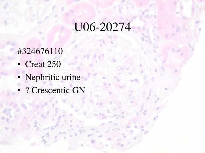
U06- 20274. #324676110 Creat 250 Nephritic urine ? Crescentic GN. 54-year old Caucasian male. CC: Leg Edema, Skin Rash, Leg Pain and hands numbness.

E N D
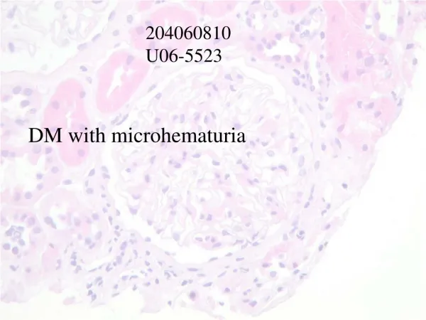
U06-20274 #324676110 • Creat 250 • Nephritic urine • ? Crescentic GN
54-year old Caucasian male • CC: Leg Edema, Skin Rash, Leg Pain and hands numbness. • HPI: 8 Wks prior to admission he noticed leg edema and rash. Treated with Cephazolin and cephalexin for few days for cellulitis. No URI, No Fever, No gross hematuria, No Sore throat. • PMH: Asthma (using combivent PRN)
Labs: (on admission) • Hgb: 132 • Cr 247 (Baseline Cr: 110) • U/A: SG of >1.030 • Pro/Crea: 314.00 • ALT, AST and Alk.Pho: NL • dsDNA: Neg, • RF(Sep2006): 22 • ANA: NEG • p-ANCA and c-ANCA: NEG X 2 • C3 and C4: 1.33 and .91
IF • IgG-Negative. • IgA- Moderate irregular peripheral lobular finely granular staining. • IgM-Negative. • C3- Moderate irregular peripheral lobular staining. Moderate vascular staining. • C1q-Negative. • Kappa-Negative. • Lambda-Negative. • Fibrin- Mild interstitial staining. • Albumin-Negative.
EM • Will be ready in the coming weeks.
DiagnosisRenal Biopsy: • Focal proliferative GN with peripheral lobular granular staining for IgA and possible arteriolitis with necrotizing changes. • Rule out Henoch-Schönlein purpura. Comment: the IF pattern is unusual but has been described in Henoch-Schönlein purpura.
Series of 10 paediatric cases • A/a focal and segmental hypercellularity with # monocytes • IgA deposits at periphery of lobules, not in mesangium • Early phase of disease ? • 3/6 subsequent biopsy was normal (with no IgA deposition) • 3/6 subsequent biopsy with mesangial IgA