Download
1 / 140
1.41k likes | 1.64k Views
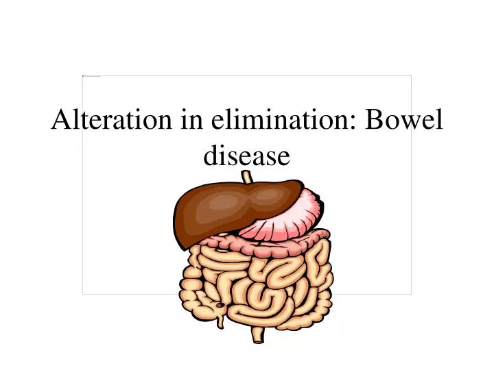
Alteration in elimination: Bowel disease. Alteration in elimination bowel. Inflammatory bowel disease Small bowel obstruction Cancer of the colon and ostomies. Small intestine. Made up of three parts: ileum, jejunum, and duodenum. Main function is absorption. Small intestine.

E N D
Alteration in elimination bowel • Inflammatory bowel disease • Small bowel obstruction • Cancer of the colon and ostomies.
Small intestine • Made up of three parts: ileum, jejunum, and duodenum. • Main function is absorption
It is a symptom not a primary disorder. It is the increase in: fluid, volume, and fluid content of the stool. Causes: Bacteria toxins Parasitic infections Malabsorption syndromes Medication Systemic disease Allergies Psychogenic Diarrhea
Constipation • Two or less BM’s weekly or when defecation is excessively difficult or requires straining. • Most common cause: Ignoring the urge to defecate. Treat this cause with education ( a daily BM is not necessary for good health) exercise and diet modification.
Nursing assessment • Questions ask? • Have you been out of the country? • What medications have you used? • When did the diarrhea start? • Are there any associated symptoms?
Nursing assessment • Observe the patient’s stool for steatorrhea, blood, pus, or mucus. • Monitor frequency and characteristics of bowel movement. • Measure abdominal girth and auscultate bowel sounds every shift.
Nursing diagnosis • Fluid volume deficit • Risk for impaired skin integrity • Altered nutrition: less than body requirements related to loss of nutrients
Fluid and electrolyte imbalance r/t diarrhea • The increased water content of the stool places the patient at risk for fluid deficit. • Record Accurate I&O • Weight patient QD • Assess the patient’s mucous membrane, skin turgor, and urine specific gravity. • Monitor and record vital signs including orthostatic blood pressures.
Fluid and electrolyte imbalance r/t diarrhea • Postural (orthostatic) blood pressure changes. • When the BP drops more than 10mmHg when changing positions (lying to sitting, sitting to standing). Orthostatic changes indicate fluid deficit. Pulse typically increases at the same time.
Risk for impaired skin integrity • Provide good skin care • Assist the client with cleaning the perianal area as needed. Use warm water and soft cloths. • Provide protective ointment to the perianal area
Caution on pharmacological treatments • Laxatives should never be administer to a patient with bowel obstruction or impaction. • People with abdominal pain of undetermined cause. • Laxatives can cause mechanical damage and perforate the bowel.
Caution on pharmacological treatments • Enemas are use for chronic constipation or fecal impaction. • As a general rule use only for acute phase on a short time bases. • Excessive use of enema can lead to fluid electrolyte imbalance. • Never use enemas if you suspect perforation.
Chronic inflammatory bowel disease • Two inflammatory diseases(Crohn’s disease and Ulcerative colitis )similar on the following : • Etiology is unknown (autoimmune component involve) • genetic components/run families/ethnic groups • Affect young adults between the ages 15-35 years. • Diarrhea is the predominant symptom
Ulcerative colitis • Affects the mucous and the submucosa of the colon and rectum. • Primarily affects the young (15-30) • More common in whites • Cause unknown found in families with hx. of the same, hx crohn’s, Hx certain arthritis.
Pathophysiology of ulcerative colitis • Inflamed crypts of Lieberkuhn in the distal large intestine and rectum • Pinpoint microscopic hemorrhages develop • Then crypt abscesses develop. • The abscesses penetrated the superficial submucosa an spread laterally leading to mucosal necrosis and sloughing.
Pathophysiology of Ulcerative Colitis • The inflammatory process leads to further tissue damage from exudate and the release of inflammatory mediators such as prostanglandins and cytokines. • The mucosa becomes red because of vascular congestion, friable and edematous. • It bleeds easy and hemorrhage is common.
Pathophysiology of Ulcerative Colitis • Edema obscure the submucosal vessels and creates a granular appearance. • Pseudopolyps tongue line projections are common. • Polypoid changes represent areas of edematous tissue between areas of ulceration .. • Chronic inflammation leads to shortening of the colon from fibrosis and loss of haustra.
Pathophysiology of Ulcerative Colitis • The inflammatory process begins at the rectosigmoid are of the anal canal and progresses proximal. • May progress to involve the entire colon. • Blood, mucus and pus pool in he lumen of the colon (characteristic diarrhea) • The extent of the colon involving correlates with severity of the disease.
Ulcerative Colitis signs and symptoms • Insidious onset • Attacks last 1-3 months • Occur at intervals of months to years • Diarrhea is the predominant symptoms of all types of ulcerative colitis. • Typically 30-40 stools per day, with blood and mucus.
Ulcerative Colitis signs and symptoms • When severe disease is present may have other manifestation such as arthritis (related to the inflammatory process going on), uveitis, thromboemboli, lesions of the liver, gallbladder, and pancreas as well as pericarditis. • Patients with Ulcerative Colitis have an increased risk of developing colon cancer.
Complications of Ulcerative Colitis • Bowel perforation most deadly • Hemorrhage • Toxic megacolon • Increased risk of developing colon cancer. • The risk is higher when there is intensive involvement of the colon with disease for >10 years.
Diagnostic of Ulcerative Colitis • Stool for occult blood • Hemoglobin and hematocrit • Colonoscopy**not on active phase • Barium enema**” • A yearly colonoscopy is strongly recommended for anyone who has ulcerative colitis with 8-10years after the DX.
Treatment of Ulcerative Colitis • Pharmacological • Dietary management • Surgical management
Pharmacological treatment • Sulfasalazine (Azulfidine) anti-inflamatory • inhibits prostaglandin production in the bowel. • Mesalamine (Rowasa) & Olsalazine (Dipentum) -Same action as above. • Corticosteroids-anti-inflammatory effects • Use as a treatment during acute attacks.
Immunossupression Imuran (Azathioprine) Cyclosprine (Sandimmune) Antidiarrheal (not used during an acute attack) Loperamide Diphenoxylate Pharmacological treatment
Dietary management in ulcerative colitis • No milk products • No caffeine • No gas producing or raw fruits & vegetables • Bulk forming products such as psyllium or methylcellulose to decreased diarrhea and reduce symptoms. • TPN during acute exacerbation
Surgery as a treatment for ulcerative colitis. • Procedure of choice is a total colectomy with ileonal anastomosis. • The entire colon and rectum are remove • A pouch is formed from the terminal ileum
Surgery as a treatment for ulcerative colitis. • The pouch is brought into the pelvis and anastomosed to the anal canal. • A temporary or loop ileostomy is performed and maintained for 2 to 3 months. • When the anastomosis sites heal the ileostomy is closed and the patient has bowel movements through the anus.
Surgery as a treatment for ulcerative colitis. • The Kock’s ileostomy(continent) • an intra-abdominal reservoir is constructed from the terminal ileum. • Stool collects in the pouch until the patient drains it with a catheter • A nipple valve prevent leakage of stool.
Surgery as a treatment for ulcerative colitis. • Total proctocolectomy with permanent ileostomy. • Colon, rectum, and anus are remove, and the end of the terminal ileum is exteriorized as a stoma on the right abdominal wall.
Surgery as a treatment for ulcerative colitis. • Temporary or loop ileostomy is often used to eliminate feces and allow healing for 2-3 months after an ileoanal anastomosis. • A loop of the ileum is brought to the body surface and allows stool drainage into the external pouch. • When the stoma is not needed a second surgery is done to close the stoma and repair the bowel. • See Lemone text pp.826-829 for nursing care of patients with an ileostomy, for changing an ostomy pouch, and for ileostomy lavage.
Relieving abdominal cramping Providing emotional support Teaching about the illness and special needs. Nursing diagnosis: Fluid and electrolytes imbalance R/T diarrhea Body image disturbance R/T disease process Nursing care in ulcerative colitis
Monitor the appearance and frequency of bowel movement. Assess and document presence of blood in the stool by testing for occult blood and BRB Assess document Vital signs q4hrs. Record pt. wt. qd. Assess the pt. for signs of fluid deficit. Maintain fluid intake by mouth or by parenteral means as indicated Fluid and electrolyte imbalance