Download
1 / 37
370 likes | 532 Views
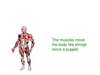
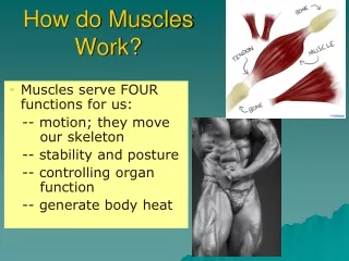
chapter. 1. MUSCLES AND HOW THEY MOVE. Learning Objectives. w Learn the basic components of skeletal muscle , the muscle fiber , and the myofibril. w Note the cellular events leading to a basic muscle action. w Discover how muscle functions during exercise.

E N D
chapter 1 MUSCLES AND HOW THEY MOVE
Learning Objectives w Learn the basic components of skeletal muscle, the muscle fiber, and the myofibril. w Note the cellular events leading to a basic muscle action. w Discover how muscle functions during exercise. w Consider the differences in fiber types and their impact on physical performance. w Learn how muscles generate force and movement by pulling on bones.
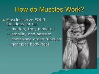
Types of Muscles • Skeletal • w Voluntary muscle; controlled consciously • Over 600 throughout the body Cardiac w Controls itself with assistance from the nervous and endocrine systems w Only in the heart • Smooth • w Involuntary muscle; controlled unconsciously • In the walls of blood vessels and internal organs
Key Points Muscle Fiber w An individual muscle cell is called a muscle fiber. w A muscle fiber is enclosed by a plasma membrane called the sarcolemma肌漿膜. w The cytoplasm of a muscle fiber is called the sarcoplasm. w Within the sarcoplasm, the T tubules allow transport of substances throughout the muscle fiber. wThe sarcoplasmic reticulum stores calcium.
Key Points Myofibrils w Myofibrils are the contractile elements of skeletal muscle, with several hundred to several thousand composing a single muscle. w Myofibrils are made up of sarcomeres, the smallest functional units of a muscle. w A sarcomere is composed of filaments of two proteins, myosin and actin, which are responsible for muscle contraction. wMyosin is a thick filament with a globular head at one end. w An actin filament—composed of actin, tropomyosin, and troponin—is attached to a Z disk.
5. The Ca2+ binds to troponin on the actin filament, and the troponin pulls tropomyosinoff the active sites, allowing myosin heads to attach to the actin filament. (continued) Excitation/Contraction Coupling 1. A motor neuron, with signals from the brain or spinal cord, releases the neurotransmitter acetylcholine (Ach) at the neuromuscular junction. 2. ACh crosses the junction and binds to receptors on the sarcolemma. 3. This initiates an action potential, providing sufficient ACh. 4. The action potential travels along the sarcolemmaand through the T tubules to the SR releasing Ca2+.
Excitation/Contraction Coupling 6. Once a strong binding state is extablished with actin, the myosin head tilts, pulling the actin filament (power stroke). 7. The myosin head binds to ATP, and ATPase found on the head splits ATP into ADP and Pi, releasing energy. 8. Muscle action ends when calcium is actively pumped out of the sarcoplasm back into the sarcoplasmic reticulum for storage.fig1.9
Excitation/Contraction Coupling Fig 1.8 a sarcomere in its relaxed and contracted state(I,A band and H zone) Fig 1.9 change in the myosin head during various phases of the power stroke(number 1 and 2)
Sliding Filament Theory w When myosin cross-bridges are activated, they bind strongly with actin, resulting in a change in the cross-bridge. w The change in the cross-bridge causes the myosin head to tilt toward the arm of the cross-bridge and drag the actin and myosin filaments in opposite directions. w The tilt of the myosin head is known as a power stroke. w The pulling of the actin filament past the myosin results in muscle shortening and generation of muscle force.
Muscle Biopsy w Hollow needle is inserted into muscle to take a sample. w Sample is mounted框好, frozen, thinly sliced, and examined under a microscope. w Allows study of muscle fibers and the effects of acute exercise and exercise training on fiber composition.
Slow-Twitch (St,typeⅠ) Muscle Fibers wHighaerobic (oxidative) capacity and fatigue resistance wLow anaerobic (glycolytic) capacity and motor unit strength wSlow contractile speed (110 ms to reach peak tension) and myosin ATPase w 10–180 fibers per motor neuron wLow sarcoplasmic reticulum development
Fast-Twitch (Fta,typeⅡa) Muscle Fibers wModerate aerobic (oxidative) capacity and fatigue resistance wHigh anaerobic (glycolytic) capacity and motor unit strength wFast contractile speed (50 ms to reach peak tension) and myosin ATPase w 300–800 fibers per motor neuron wHigh sarcoplasmic reticulum development
Fast-Twitch (FTx,type Ⅱx) Muscle Fibers wLow aerobic (oxidative) capacity and fatigue resistance wHigh anaerobic (glycolytic) capacity and motor unit strength wFast contractile speed (50 ms to reach peak tension) and myosin ATPase w 300–800 fibers per motor neuron wHigh sarcoplasmic reticulum development
Did You Know…? The difference in force development between FT and ST motor units is due to the number of muscle fibers per motor unit and the larger diameter of the FT fibers. Table 1.1 and table 1.2.
What Determines Fiber Type? wGenetics determine which type of motor neurons innervate our individual muscle fibers. w Muscle fibers become specialized according to the type of neuron that stimulates them. w Endurance training, strength training, and muscular inactivity may result in small changes (less than 10%) in the percentage of FT and ST fibers. wEndurance training has been shown to reduce the percentage of FTx fibers, while increasing the fraction of FTa fibers. • Aging may result in changes in the percentage of FT and ST fibers.
wFT fibers have a more highly developed sarcoplasmic reticulum enhancing calcium delivery. (continued) Key Points Slow- and Fast-Twitch Muscle Fibers w Skeletal muscles contain both ST and FT fibers. w ATPase in FT fibers acts faster providing energy for muscle action more quickly than ATPase in ST fibers.
Key Points Slow- and Fast-Twitch Muscle Fibers w Motor units supplying FT fibers are larger (e.g., more fibers per motor neuron) than those supplying ST fibers; thus, FT motor units can recruit more fibers. wST fibers have high aerobic endurance and are suited to low-intensity endurance activities. wFT fibers are better for anaerobic or explosive activities.
All-Or-None-Response w For a motor unit to be recruited into activity the motor nerve impulse must meet or exceed the threshold. w When this occurs, all muscle fibers in the motor unit act maximally. w If the threshold is not met no fibers in that unit act. wMore force is produced by activating more motor units.
Orderly Recruitment of Muscle Fibers • Principle of orderly recruitment states that motor units are activated in a fixed order, based on their ranking in the muscle. • Slow-twitch fibers, which have smaller motor neurons, are recruited before fast-twitch fibers.
Table 1.3 percentage and cross-sectional areas of typeⅠand typeⅡ fiber.
Factors Influencing Force Generation w Number of motor units and muscle size(type2>type1) w Frequency of stimulation of the motor units fig1.12 wSarcomere length fig1.13(b and c peak tension) wSpeed of muscle action (shortening or lengthening) fig1.14