Download
1 / 1
20 likes | 101 Views
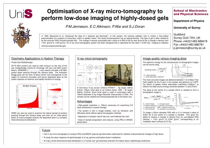
S. Dosimetry Applications in Hadron Therapy Proton Eye Radiotherapy:

E N D
S Dosimetry Applications in Hadron Therapy Proton Eye Radiotherapy: 50-70 MeV protons are used to treat tumours on the rear of the eye (Clatterbridge Centre for Oncology, UK has a 62 MeV proton beam). SRIM2003 (www.srim.org) was used to simulate a 50 MeV proton beam passing through the vitreous body. The ionisation Bragg peak can be seen at about 22mm and corresponds to the region of maximum ionisation and hence deposited dose as the proton captures an electron and rapidly transfers its energy. SRIM can also be used to examine the lateral spread of protons passing through the vitreous body and even for an initial pencil beam of mono-energetic protons the deposited dose is a complex three-dimensional distribution. Optimisation of X-ray micro-tomography to perform low-dose imaging of highly-dosed gels School of Electronics and Physical Sciences Department of Physics University of Surrey Guildford Surrey GU2 7XH, UK Phone+44(0)1483 689419 Fax+44(0)1483 686781 p.jenneson@surrey.ac.uk P.M.Jenneson, E.C.Atkinson, P.Wai and S.J.Doran In 1993, Maryanski et al. introduced the idea of a "polymer gel dosimeter". In this system, the ionising radiation sets in motion a free-radical polymerisation of a solution of monomers, held in a gelatin matrix. The newly formed polymer has an altered density. This type of gel is often refered to by the acronym PAG (PolyAcrylamide Gel). The change in density associated with a dose to a PAG of 50 Gy was found to be a change in density from 1.021 g/cm3 to 1.035 g/cm3. An X-ray micro-tomography system has been designed that is optimised for low dose (~0.054 Gy) imaging of radiation sensitive polyacrylamide gels. • X-ray micro-tomography • A mini-focus X-ray source (Oxford XTF5011 :- Ag target, 50kVp 0.5mA, 100um focal spot or an Oxford series 1500 :- W target, 80kVp, 0.25mA, 33um focal spot) is used in conjunction with a 100mm diameter X-ray image intensifier (Hamamatsu C7336). The sample can be rotated and moved perpendicular to the beam axis. • Advantages • High-spatial resolution (< 100um) necessary for examining 3-D dose distributions in proton therapy. • The system is low in image artefact, such as refraction and diffraction which affects optical tomographic methods • Apparatus is compact, bench top size, and relatively low cost • Ease of sample preparation and analysis, using PAG or MAGIC dosemeters. Image quality versus imaging dose The optimum energy for the reconstruction of tomographic images can be found using, D = object diameter = linear attenuation coefficient The most accurate images are obtained between 1 to 6 Mean Free Path lengths for the X-rays in the sample and optimum at 3 MFP. For 25mm of gelatin (linear attenuation coefficient 0.656cm-1 at 22keV) the ideal source energy would be between 13 and 27keV. The dose to the centre of a sample which is needed to form a tomographic image is To image a 25mm diameter PAG with a 0.1% contrast a dose of 0.054 Gy to the centre of a sample is needed. This gives the ability to resolve a change in density from an unirradiated PAG (density = 1.021 g/cm3) of 0.001 g/cm3, corresponding to an irradiation dose contrast of 3.5 Gy. Ex= beam energy • = detector efficiency • = plane pixel size h = slice thickness • fc = Compton factor • = density of the sample SNR = signal-to-noise ratio d = diameter of reconstruction • Future • Use X-ray micro-tomography to analyse PAG and MAGIC polymer gel dosimeters optimised for radiation induced density changes at high doses • To study the dose response of optimised gels to X-ray, gamma and hadron beam irradiations • To map a three-dimensional dose distribution in a “human eye” gel dosimeter phantom for hadron beam radiotherapy treatments