Download
1 / 68
680 likes | 1.21k Views
Overview. History of stapes surgeryCauses of stapes fixationReview of otosclerosisPatient evaluationStapes surgical techniqueComplications of stapes surgeryIntraoperativePost-operative. History of Stapes Surgery. Samuel Rosen1953

E N D
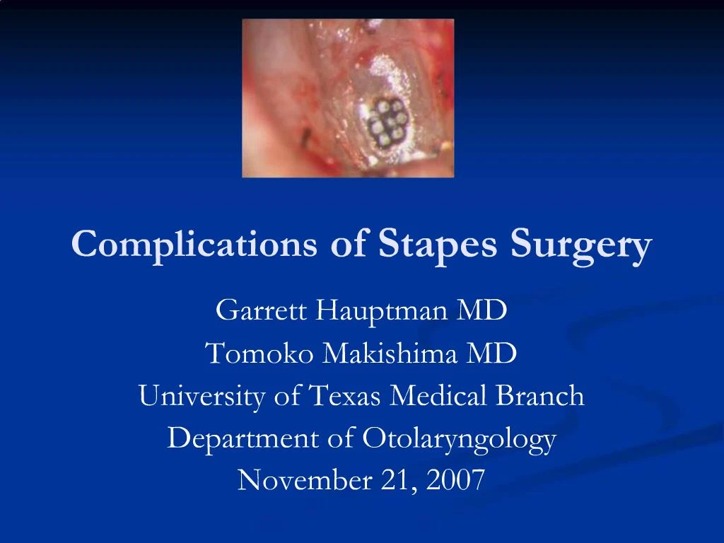
1. Complications of Stapes Surgery Garrett Hauptman MD
Tomoko Makishima MD
University of Texas Medical Branch
Department of Otolaryngology
November 21, 2007
2. Overview History of stapes surgery
Causes of stapes fixation
Review of otosclerosis
Patient evaluation
Stapes surgical technique
Complications of stapes surgery
Intraoperative
Post-operative
3. History of Stapes Surgery Samuel Rosen
1953 � first suggest mobilization of the stapes
Immediately improved hearing
Problem with re-fixation
4. History of Stapes Surgery John Shea
1956 � first to perform stapedectomy
Oval window vein graft
Nylon prosthesis from incus to oval window
5. Overview History of stapes surgery
Causes of stapes fixation
Review of otosclerosis
Patient evaluation
Stapes surgical technique
Complications of stapes surgery
Intra-operative
Post-operative
6. Causes of Stapes Fixation Otosclerosis
= 95% of stapes surgery
Congenital stapes fixation
Hearing outcomes worse with stapes surgery compared to otosclerosis
Groups stratified into ABG < 10 db and ABG < 20 dB
Tympanosclerosis
Hearing outcomes worse with stapes surgery compared to otosclerosis
Mobilization through plaque removal �vs- stapedotomy
7. Overview History of stapes surgery
Causes of stapes fixation
Review of otosclerosis
Patient evaluation
Stapes surgical technique
Complications of stapes surgery
Intraoperative
Post-operative
8. Otosclerosis Bone disease only seen in otic capsule
Causes progressive hearing loss
Conductive- primarily stapes involvement
Sensorineural- cochlear involvement
Mixed
9. Epidemiology
10% overall prevalence of histologic otosclerosis
1% overall prevalence of clinically significant otosclerosis
Bilaterality more common
10. Epidemiology
Race Incidence of otosclerosis
Caucasian 10%
Asian 5%
African American 1%
Native American 0%
11. Epidemiology Gender
Histologic otosclerosis � 1:1 ratio
Clinical otosclerosis � 2:1 (W:M)
Possible progression during pregnancy (10%-17%)
Studies demonstrating changes during pregnancy usually retrospective or lack audiometric data
Studies comparing multigravid �vs- nulligravid women with otosclerosis fail to show audiometric differences
12. Epidemiology Age
15-45 most common age range of presentation
Youngest presentation 7 years
Oldest presentation 50s
0.6% of individuals < 5 years old have foci of otosclerosis
13. Pathophysiology of Otosclerosis
Osseous dyscrasia
Resorption and formation of new bone
Limited to the temporal bone and ossicles
Inciting event unknown
Hereditary, endocrine, metabolic, infectious, vascular, autoimmune, hormonal
14. Pathology Two phases of disease
Active (otospongiosis phase)
Osteocytes, histiocytes, osteoblasts
Active resorption of bone
Dilation of vessels
Schwartze�s sign
Mature (sclerotic phase)
Deposition of new bone (sclerotic and less dense than normal bone)
15. Pathology
Most common sites of involvement
Fissula ante fenestrum
Round window niche (30%-50% of cases)
Anterior wall of the IAC
17. Overview History of stapes surgery
Causes of stapes fixation
Review of otosclerosis
Patient evaluation
Stapes surgical technique
Complications of stapes surgery
Intraoperative
Post-operative
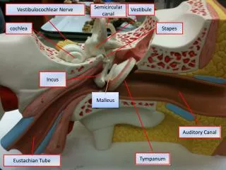
18. Patient Evaluation History
Gradual onset with slow progression over several years
Typically presents during late teens or twenties
70% are bilateral
Family history usually positive
19. Patient Evaluation Physical examination
Otoscopy (often with the operating microscope)
look for Schwartze sign: red blush over the promontory or area anterior to oval window
Pneumo-otoscopy
evaluates for middle ear effusion or small perforation
Tuning fork exam
may confirm or dispute finding of conductive hearing loss on audiometry
20. Patient Evaluation Audiometry
Standard audiometry
Air conduction
Bone conduction
Speech audiometry
Immittance audiometry
Tympanometry- lower peak than normal (As)
Static compliance
Acoustic reflexes- absent in advanced disease
21. Overview History of stapes surgery
Causes of stapes fixation
Review of otosclerosis
Patient evaluation
Stapes surgical technique
Complications of stapes surgery
Intraoperative
Post-operative
22. Stapes Surgery Informed consent
Total sensorineural hearing loss occurs 0.2% of cases
Less than 2% chance of further hearing loss
Dizziness may occur post-operatively
Usually transient and brief
May persist for short time
Rarely could be permanent
Possible facial paralysis/palsy
Tinnitus
Recurrent conductive hearing loss
23. Middle Ear Examination Mobility of ossicles
Confirm stapes fixation
Evaluate for malleus or incus fixation
Abnormal anatomy
Dehiscent/overhanging facial nerve
Deep narrow oval window niche
Ossicular abnormalities
24. Stapedectomy �vs- Stapedotomy Stapedectomy
Uses
Extensive fixation of the footplate
Floating footplate
Disadvantages
Increased post-op vestibular symptoms
More technically difficult
Increased potential for prosthesis migration
Stapedotomy
Originally for obliterated or solid footplates
Europe
1970-80
First laser stapedotomy performed by Perkins (1978)
Less trauma to the vestibule
Less incidence of prosthesis migration
Less fixation of prosthesis by scar tissue
25. Stapedotomy Microdrill
0.7mm diamond burr
Motion of the burr removes bone dust
Minimizes smoke production/surrounding heat production
Laser
Avoids manipulation of the footplate
Argon and Potassium titanyl phosphate (KTP/532)
Wave length 500 nm
Visible light
Absorbed by hemoglobin
Surgical and aiming beam
Carbon dioxide (CO2)
10,000 nm
Not in visible light range
Surgical beam only
Requires separate laser for an aiming beam (red helium-neon)
26. Stapedectomy �vs- Stapedotomy ABG closure < 10dB (PTA)
27. Sequence of Stapes Surgery Retrospective review
376 patients
420 stapedotomies
Measured incidence of:
Incus subluxation
Floating footplate
Results
Footplate perforation before stapes arch removal ? risk of floating footplate
Incus subluxation ? when prosthesis placed prior to stapes arch removal
28. Classic Stapes Surgery Approach Stapes superstructure removed
Fenestration of footplate
Prosthesis placement
29. Modified Stapes Surgical Approach Fenestration of footplate
Stapes superstructure removal
Prosthesis placement
30. Modified Stapes Surgical Approach Fenestration of footplate
Prosthesis placement
Stapes superstructure removal
31. Overview History of stapes surgery
Causes of stapes fixation
Review of otosclerosis
Patient evaluation
Stapes surgical technique
Complications of stapes surgery
Intra-operative
Post-operative
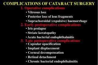
32. Problems During Stapes Surgery Exposed overhanging facial nerve
Occurs ~9% of stapes procedures
May block footplate access making completion impossible
Prosthesis touching facial nerve generally does not create problem
May displace nerve superiorly while performing stapedotomy
33. Problems During Stapes Surgery Floating Footplate
Footplate dislodges from surrounding oval window niche
Usually iatrogenic
Incidental finding
Prevention
Laser
Footplate control hole
Management
Abort
Proceed
Total stapedectomy
Laser fenestration/microdrill fenestration
34. Problems During Stapes Surgery Diffuse Obliterative Otosclerosis
Occurs when footplate, annular ligament, and oval window niche are involved
Closure of air-bone gap < 10 dB less common
Refixation commonly occurs
Fenestra created with microdrill
35. Problems During Stapes Surgery Fixed malleus
Rare problem
Must always check
Must check mobility of prosthesis after placement
36. Problems During Stapes Surgery Perilymph Gusher - profuse flow of perilymph immediately upon opening vestibule
Rare � 0.03% incidence
Associated with congenital footplate fixation
Possibly due to:
Widened vestibular aqueduct
Defect in IAC fundus
Management
Tissue graft over oval window
Complete procedure if possible
Consider lumbar drain
37. Problems During Stapes Surgery Intraoperative vertigo
Causes
Prosthesis too long
Checking prosthesis mobility
Management
Shorter prosthesis (try 0.25mm shorter piston)
38. Post-operative Complications Sensorineural Hearing Loss
Most devastating complication of stapes surgery
Ranges from mild to total loss or may be isolated to high frequencies
<1% - 3% incidence of profound permanent SNHL
Surgeon experience
Extent of disease
Cochlear
Prior stapes surgery
39. Post-operative Complications Sensorineural Hearing Loss (cont.)
Temporary
Serous labyrinthitis
Reparative granuloma
Permanent
Suppurative labyrinthitis
Extensive drilling
Basilar membrane breaks
Vascular compromise
Sudden drop in perilymph pressure
Management
Prednisone taper started immediately
40. Post-operative Complications Sensorineural Hearing Loss (cont.)
Prospective study with Otology-Neurotology Database
3050 stapedotomies for otosclerotic stapes fixation (2525 patients)
Results
Significant post-op SNHL (> 15dB)
0.5% overall
4.8% in obliterative otosclerosis
0 cases with simultaneous malleus ankylosis
41. Post-operative Complications Recurrent Conductive Hearing Loss
Slippage or displacement of the prosthesis
Most common cause of failure
Immediate
Technique
Trauma
Delayed
Slippage from incus narrowing or erosion
Adherence to edge of oval window niche
Stapes re-fixation
Progression of disease with re-obliteration of oval window
Malleus or incus ankylosis
42. Post-operative Complications Recurrent Conductive Hearing Loss (cont.)
Prospective study
260 pts with ABG = 20dB after stapedotomy or stapedectomy
1 month to 35 years after surgery
Cause of CHL
81% prosthesis displacement
Other causes:
Residual footplate fixation
Malleus fixation
Incus fixation
Incus dislocation
43. Post-operative Complications Recurrent Conductive Hearing Loss (cont.)
Recommendations
Laser stapedotomy
Teflon/platinum stapedotomy prosthesis
Prosthesis 0.25mm longer than distance between incus undersurface and footplate
Clotted blood oval window seal
Minimize mechanical trauma
Use tissue seal
Perilymph gusher
Footplate fracture
When stapedotomy too large
44. Conductive Hearing Loss Mechanism: After Stapedotomy Collagen tissue seal contracts
Prosthesis lifts out of stapedotomy
Prosthesis migrates to fixed stapes footplate
45. Conductive Hearing Loss Mechanism: After Stapedectomy Collagen tissue seal contracts
Neomembrane lateralizes
Erosion of incus causing loosening of wire loop
46. Post-operative Complications Serous labyrinthitis
Common following surgery secondary to inner ear inflammation
Symptoms
Unsteadiness
Positional vertigo
Slight high frequency hearing loss
Management
Expectant
47. Post-operative Complications Vertigo
More common with stapedectomy than stapedotomy
Due to serous labyrinthits
Occurs ~5% of cases
Rarely prolonged or severe
Usually lasts a few hours to one week
Rapidly subsides
Supportive management
48. Post-operative Complications Vertigo (cont.)
Intraoperative or immediately post-op: lasts up to 1 week without intervention
Inner ear trauma
Prosthesis/instrument contact with membranous labyrinth (utricular macula)
Perilymph aspiration
Isolated delayed vertigo
Trauma to otolith organs creating BPPV-like picture
Perilymphatic fistula
49. Post-operative Complications Delayed Vertigo
Retrospective review
9 pts with delayed vertigo (1month to seven years post-op) underwent exploratory tympanotomy
Suspected perilymph fistula in all pts
3 pts had perilymph fistula
Fibrin glue placed in oval window area in all pts
No post-operative vertigo
50. Post-operative Complications Perilymph Fistula
Rare complication after stapes surgery
Presents with:
Mixed hearing loss
Vague unsteadiness
Vertigo
Management
Remove prosthesis carefully ? tissue seal the oval window ? prosthesis replaced
51. Mechanism of Post-operative Perilymph Fistula: Stapedotomy Incus medially displaced by contracture adhesions between incus and promontory
Prosthesis medializes into vestibule
52. Mechanism of Post-operative Perilymph Fistula: Stapedectomy Prosthesis migration from center to edge of oval window
Vibration tears weaker shortened edge of membrane
53. Post-operative Complications Tinnitus
Possibly related to serous labyrinthitis
Management
Reassurance
Routine tinnitus measures
54. Post-operative Complications Facial paralysis/palsy
Rare
Delayed onset
Typically lasts several weeks
Occurs in 5-day post-op setting
Usually incomplete paralysis
Management
Prednisone- usually complete response
55. Post-operative Complications Facial paralysis/palsy (cont.)
Retrospective review
2152 stapes surgeries (2106 pts)
0.51% delayed facial palsy
Occurred 5-16 days post-op
Measurements
House-Brackmann grade
Serum antibody titer (HSV1, HSV2, VZV)
Conclusion
Serology suggests activation of latent herpesvirus
56. Post-operative Complications Facial paralysis/palsy (cont.)
Retrospective review
706 stapes surgeries (580 pts)
0.01% delayed facial palsy
Measurements
House-Brackmann grade
Serum antibody titer (HSV1)
Conclusion
Serology suggests activation of latent herpesvirus
Treat with acyclovir
57. Post-operative Complications Reparative granuloma
Very rare- associated with Gelfoam use
Patient presentation
Initial hearing improvement followed by gradual/sudden deterioration over 1 to 6 weeks
Reddish discoloration in posterosuperior quadrant
Occasional vertigo
Management
Granuloma removal
58. Post-operative Complications Chorda Tympani damage
Occurs ~30% of cases due to nerve stretching/mobilization
Causes temporary (3-4 months)
Dry mouth
Tongue soreness
Metallic taste
Symptoms less severe with sectioning of nerve
59. Post-operative Complications Tympanic membrane perforation
May occur during elevation of tympanomeatal flap
Does not preclude completion of operation
Repair involves myringoplasty or tympanoplasty with either synthetic material or autologous tissue
60. Post-operative Complications Meningitis
Creation of fistula introduces route for potential meningitis
Case report
33yo? POD 1 with vertigo, n/v, hearing loss, severe pain
Later developed neck stiffness
LP with cloudy CSF
Blood Cx with streptococcus pneumoniae
Treated with IV antibiotics
61. Post-operative Complications Psychiatric complication
Case report
Underlying schizoaffective disorder
Stapedectomy performed with complete closure of ABG
Pt believed surgery resulted in:
Improved sound perception
Thought broadcasting
62. Prosthesis Selection Robinson piston
Relatively heavy � may increase risk of displacement into vestibule
Handle can cause necrosis
Wire piston
Incus necrosis due to:
mass
crimping tightness
Crimping angle may favor movement resulting in displacement over time
63. Prosthesis Selection Vertigo assessment
Randomized-blinded controlled trial
174 original Fisch prosthesis �vs- 108 modified prosthesis
No difference in closure of ABG
Post-operative vertigo reduced
64. Modified Prosthesis 45 degree slope at distal piston end
Anatomically configured to avoid saccule
65. Revision Stapes Surgery Retrospective review
63 surgeries (56 pts)
Revision reason
Recurrent or persistent ABG > 20dB post-surgical treatment for otosclerosis
Prosthesis malfunction was primary failure cause
66. Revision Stapes Surgery Results
52.4% ABG = 10 dB
9.5% without change
6.3% decreased hearing = 5 dB
Recommendations
Examine
Prosthesis attachment to incus
Oval window niche
Pistons can be removed easily
Tissue wire prostheses
Difficult to remove- laser helps with removal
Increased risk of SNHL
67. Stapes Surgery by Residents Retrospective review
71 stapedotomies (laser-assisted fenestra)
87% with closure of air-bone gap = 10 dB
Complications
High-frequency SNHL of 15-30 dB in 3 pts
Transient vertigo in 3 pts
No sensorineural deafness
68. Conclusion Stapes surgery
Delicate structures
Small area
Important surroundings
Surgeon must be aware of potential complications and management
Informed consent is essential
69. Bibliography Albera R et al. Delayed vertigo after stapes surgery. Laryngoscope 2004; 114: 860-2.
Cummings CW. Otolaryngology: Head and Neck Surgery 4th edition. Chapter 156; 2005.
Gros A et al. Success rate in revision stapes surgery for otosclerosis. Otol Neurotol 2005; 26: 1143-8.
Lesinski SG. Causes of conductive hearing loss after stapedectomy or stapedotomy: a prospective study of 279 consecutive surgical revisions. Otol Neurotol 2002; 23: 281-8.
Mangham CA. Platinum ribbon-Teflon piston reduces device failure after stapes surgery. Otolaryngol Head Neck Surg 2000; 123: 108-13.
Massey BL et al. Stapedectomy in congenital stapes fixation: are hearing outcomes poorer? Otolaryngol Head Neck Surg 2006; 134: 816-8.
Matthews SB et al. Stapes surgery in a residency training program. Laryngoscope 1999; 109: 52-3.
Mevio E et al. Stapes surgery and psychiatric complications. Auris Nasus Larynx 2000; 27: 275-6.
Nielsen TR et al. Meningitis following stapedotomy: a rare and early complication. J Laryngol Otol 2000; 114: 781-3.
Salvinelli F et al. Delayed peripheral facial palsy in the stapes surgery. Am J Otolaryngol 2004; 25: 105-8.
Shea JJ et al. Delayed facial palsy after stapedectomy. Otol Neurotol 2001; 22: 465-70.
Szymanski M et al. The influence of the sequence of surgical step on complication rates in stapedotomy. Otol Neurotol 2007; 28: 152-6.
Vincent R et al. Surgical findings and long-term hearing results in 3.050 stapedotomies for primary otosclerosis: a prospective study with the otology-neurotology database. Otol Neurotol 2006; 27: S25-47.