Download
1 / 38
390 likes | 500 Views
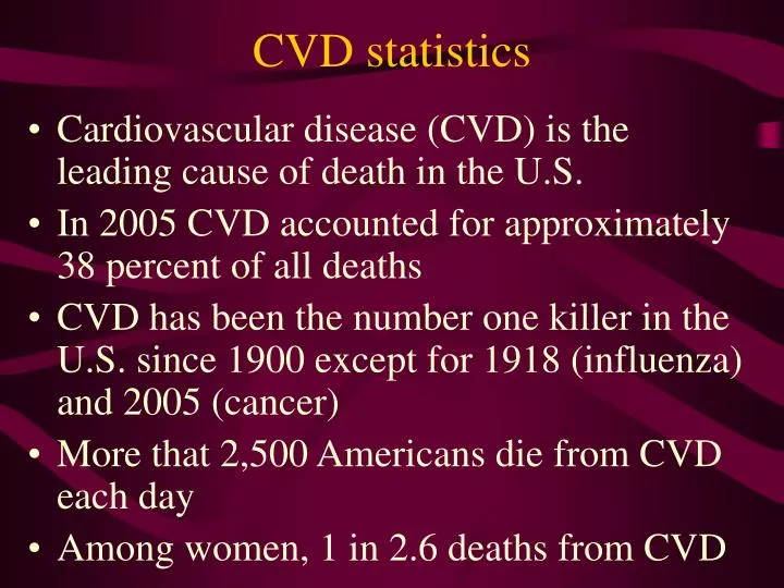
CVD statistics. Cardiovascular disease (CVD) is the leading cause of death in the U.S. In 2005 CVD accounted for approximately 38 percent of all deaths CVD has been the number one killer in the U.S. since 1900 except for 1918 (influenza) and 2005 (cancer)

E N D
CVD statistics • Cardiovascular disease (CVD) is the leading cause of death in the U.S. • In 2005 CVD accounted for approximately 38 percent of all deaths • CVD has been the number one killer in the U.S. since 1900 except for 1918 (influenza) and 2005 (cancer) • More that 2,500 Americans die from CVD each day • Among women, 1 in 2.6 deaths from CVD
Understanding the cardiovascular system • Cardiovascular system includes: the heart, arteries, arterioles, capillaries, venules, and veins • The heart • Muscular, four chambered pump • Contracts 100,000 times per day • Two upper chambers: atria • Two lower chambers: ventricles • Tricuspid, pulmonary, mitral, and aortic valves
Function of the heart • Deoxygenated blood enters the right atrium • From the right atrium blood moves to the right ventricle, pumped through the pulmonary artery to the lungs • Oxygen blood enters the left atrium • Blood from the left atrium is forced into the left ventricle • The left ventricle pumps blood through the aorta to various parts of the body
Anatomy of the Heart Figure 15.4
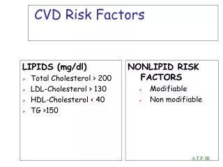
Common Types of Cardiovascular Disease • Hypertension • Atherosclerosis • Coronary heart disease (CHD) • Chest pain (angina pectoris) • Irregular heartbeat (arrhythmia) • Stroke • Congestive heart failure (CHF) • Congenital and rheumatic heart disease
hypertension High blood pressure • blood pressure- the force of blood pushing against the walls of blood vessels http://health.howstuffworks.com/adam-200079.htm
Hypertension cont’d • systolic pressure- heart contracts, sound • diastolic pressure- heart relaxes, no sound • safe range: 120/80 to 130-85; 140/90 = high
Hypertension cont’d • Called the “silent killer” because there are no symptoms and it often goes undetected
atherosclerosis arteriosclerosis- hardening of the arteries atherosclerosis- fatty deposits narrow the arteries Hyperlipidemia – abnormally high blood lipid level Plaque – the buildup of deposits in the arteries
Heart disease • Myocardial infarction (MI) or heart attack – blood supplying the heart is disrupted • Coronary thrombosis – blood clot in the artery • Embolus – when the blood clot is dislodged and moves through the circulatory system http://www.healthcentral.com/animation/408/13/Heart_Attack.html
Heart disease cont’d cardiac arrest- heart stops completely • CPR- combo. of mouth to mouth breathing and chest compressions • AED- automated external defibrillator
Heart attack cont’d Warning signs: • uncomfortable pressure/pain for two minutes or more • spreading to shoulder, neck, arms • dizziness, fainting, sweating, shortness of breath
Angina Pectoris • Ischemia – reduction of the heart’s blood and oxygen supply • The more serious the oxygen deprivation the more severe the pain
Stroke a sudden disruption of blood flow to a section of the brain Effects of Stroke
Stroke cont’d • Occurs when the blood supply to the brain is interrupted • Thrombus – blood clot • Embolus – free flowing clot • Aneurysm – bulging or burst blood vessel • Transient ischemic attack (TIA) – brief interruptions that cause temporary impairment
Stroke cont’d Warning signs: • weakness, numbness in face, arm, leg • trouble with speech • vision impairment/loss • dizziness, nausea, unsteadiness
Stroke assessment • Face weakness • Arm weakness • Speech problems • Time symptoms started
Arrhythmia • A heart condition in which the heartbeat is abnormal and irregular. • The heart may beat very slowly or very fast for no apparent reason. • Tachycardia – racing heart in the absence of exercise or anxiety • Bradycardia – abnormally slow heartbeat • Fibrillation – heart beat is sporadic, quivering pattern
Congestive Heart Failure • Damaged or overworked heart muscle is unable to keep blood circulating normally • Affects over 5 million Americans • Damage to heart muscle may result from: rheumatic fever, pneumonia, heart attack, or other cardiovascular problem • Lack of proper circulation may allow blood to accumulate in the vessels of the legs, ankles, or lungs • Diuretics relieve fluid accumulation
Detecting CVD electrocardiogram- graph of heart’s electrical activity that can show abnormalities
Detection of CVD • heart catheterization- injects dye to track blood flow and finds the blockage
Treating CVD Drugs: • Nitroglycerin – drug used to relax (dilate) the veins • Beta blockers control potential overactivity of the heart muscle
Treating CVD Aspirin Therapy • Research shows that 80 milligrams of aspirin every other day is beneficial to heart patients due to its blood thinning properties • Some side effects of aspirin: gastrointestinal intolerance and a tendency for difficulty with blood clotting • Should only be taken under the advice of your physician
Treating CVD Thrombolysis • If victim reaches an emergency room and is diagnosed quickly, thrombolysis can be performed • Thrombolysis involves injecting an agent such as tissue plasminogen activator (TPA) to dissolve the clot and restore some blood flow
Treating CVD angioplasty- balloon or laser • balloon compresses plaque against artery wall • laser dissolves plaque
Treating CVD • atherectomy- drill like catheter removes plaque
Treating CVD • Stent- wire mesh tube keeps artery wall open Stent Angioplasty
Treating CVD • coronary bypass surgery- use of a vein to construct a detour around the blocked artery Bypass Surgery
Treating CVD • pacemaker-produces electrical impulses for the heart; prevents fibrillation (uneven rhythm)
Uncontrollable Risk Factors for CVD • Increasing age • Gender • Heredity • Race
Smoking High blood cholesterol Hypertension (high blood pressure) Physical inactivity Obesity/ overweight Diabetes High stress levels Alcohol use Controllable Risk Factors for CVD
Prevention of CVD • do not smoke • if you smoke, quit • check blood pressure regularly • reduce cholesterol • reduce salt
Prevention of CVD cont’d • exercise regularly • avoid obesity • manage stress