Download
1 / 55
550 likes | 918 Views
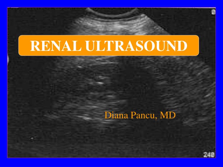
RENAL ULTRASOUND. Diana Pancu, MD. Objectives. Clinical indications for performing ED renal US Approach to performing the US study Normal anatomy Abnormal findings Clinical Impact. Clinical Indications for ED Renal Ultrasound. Suspected renal colic Colicky flank pain radiating to groin

E N D
RENAL ULTRASOUND Diana Pancu, MD
Objectives • Clinical indications for performing ED renal US • Approach to performing the US study • Normal anatomy • Abnormal findings • Clinical Impact
Clinical Indications for ED Renal Ultrasound • Suspected renal colic • Colicky flank pain radiating to groin • Hematuria • Clinical question: • Presence of hydronephrosis • Absence of other pathology (AAA)
Performing the Study • Patient preparation: • none • Transducer: 3.0MHz or 3.5 MHz • 5.0 MHz for thin patient • Patient positioning • Supine • Posterior oblique, lateral decubitus, prone
Anatomy • Kidneys are retroperitoneal, T12 - L4 • Right kidney is lower than the left kidney • Right kidney is posterio-inferior to liver & gallbladder • Left kidney is inferior-medial to the spleen • Adrenal glands are superior, anterior, medial to each kidney
Anatomy Hepatic Veins Spleen Celiac axis Liver SMA Left kidney Right kidney Renal artery Renal vein AORTA IVC
Right kidney scanning approach: anterior, lateral, posterior Liver is the acoustic window Left kidney: requires a posterior approach, through the spleen Air-filled bowel impedes anterior scanning I LIVER STOMACH SPLEEN AORTA IVC Approach to Scanning K K S
Anatomy • 9-12 cm long, 4-5 cm wide, 3-4 cm thick • Gerota’s fascia encloses kidney, capsule, perinephric fat • Sinus • Hilum: vessels, nerves, lymphatics, ureter • Pelvis: major and minor calyces • Parenchyma surrounds the sinus • Cortex: site of urine formation, contains nephrons • Medulla: contains pyramids that pass urine to minor calyces. Columns of Bertin separate pyramids
Medullary pyramids Kidney Anatomy Minor Calyx Renal artery Major Calyx Renal vein Sinus Ureter Medulla Renal capsule Cortex
Sonographic Appearance • Ureters are normally not seen • Renal pelvis is black when visible • Renal sinus is echogenic due to fat • Medullary pyramids are hypoechoic • Cortex is mid-gray, less echogenic than liver or spleen. • Capsule is smooth and echogenic
Right Kidney Long Axis Anterior Superior Inferior Liver Sinus Cortex Diaphragm Posterior
Right Kidney Short Axis Anterior Left Right GB Liver IVC R Kidney Aorta Renal a. Vertebral Body Posterior
Left Kidney Long Axis Anterior Inferior Superior Rib Shadow Kidney Posterior Spleen
Left Kidney Short Axis Anterior Right Left Liver Spleen L Kidney Posterior
Common Pitfalls in Renal Scanning • Failure to scan both kidneys • Mistaking prominent renal pyramids for hydronephrosis • Mistaking prominent pyramids for cysts • Confusing normal renal arteries for the ureter
Common Pitfalls in Renal Scanning • Failure to scan through the bladder to search for stone at the uretero-vesicular junction • Inability to visualize left kidney due to anterior probe placement • Failure to scan the aorta in suspected renal colic
Normal Variants • Dromedary humps: • Lateral kidney bulge, same echogenicity as the cortex • Hypertrophied column of Bertin: • Cortical tissue indents the renal sinus • Double collecting system: • Sinus divided by a hypertrophied column of Bertin • Horseshoe kidney: • Kidneys are connected, usually at the lower pole • Renal ectopia: • One or both kidneys outside the normal renal fossa
Clinical Indications • Obstructive Uropathy
Nephrolithiasis • 12% of the US population • Incidence of renal colic is 3% with 50% recurrence within 10 years • Manthey DE. Emerg Med Clin North Am.2001; 19(3): 633-54
Radiographic Modalities Radiography • 62% Sensitivity, 67% Specificity • Sharma RN, Shah I, Gupta S, et al: Thermogravimetric analysis of urinary stones. Br J Urol 64:564-566, 1989
Radiographic Modalities IVP vs. US • Prospective study, 85 patients • Sinclair D, Wilson S, Toi A, et al. Ann Emerg Med 18:556-559, 1989 • ULTRASOUND Sensitivity=85% • Specificity=92% • IVP • Sensitivity=90% • Specificity=94%
Radiographic Modalities ED Ultrasound + KUB vs. IVP • Prospective study, 108 patients Sensitivity = 97% Specificity = 59% Henderson, S, et al: Acad Emerg Med.1998;5:666-671. • Sensitivity = 97% • Specificity = 59% • PPV = 81% • NPV = 92%
Radiographic Modalities Helical CT- Gold Standard • Accurate, fast, no contrast • Identifies presence and size of stone • Location of stone • Level of obstruction • Other sources of pain
Stone on CT • Usually visualized • Not visualized • Stone is extremely small < 1 mm • Stone is of relatively low CT attenuation: Indinavir stones • Stone excluded from imaging due to respiratory variation
Sensitivity Ureteral dilatation 90% Perinephric stranding 82% Collecting system dilatation 83% Renal enlargement 71% Specificity Ureral dilatation 93% Perinephric stranding 93% Collecting system dilatation 94% Renal enlargement 89% Helical CTSecondary Findings Smith. AJR Am J Roentgenol 167:1109-1113, 1996
Location of Stone • 378 patients • Rate of spontaneous stone passage • 22% for proximal ureteral stones • 46% for midureteral stones • 71% for distal ureteral stones • Morse R. J Urol. 1991; 145:263-265
Width of Stone • 520 patients • Rate of spontaneous stone passage • 100% for stones that were 1 mm or smaller in width • 90% for stones 2 to 3 mm • 80% for stones that were 4 mm • 55% for stones that were 5 mm • 35% for stones that were 6 mm • 25% for stones that were 7 mm • 12% for stones that were 8 mm • Ueno A. Urology. 1977; 10:544-546
Radiographic Modalities Ultrasound • Fast • Can identify other causes of pain • Safe in pregnant patients, children
Hydronephrosis Dilatation of the urinary tract at any level secondary to intrinsic and or extrinsic obstruction to urine flow
Intrinsic, acquired Renal lithiasis Neoplasm (renal, ureteral, bladder) Papillary necrosis Ureterocele Blood clot Neurogenic bladder Anticholinergics Pregnancy, PID, uterine prolapse) Diuretics Vesico-ureteral reflux Diabetes insipidus Intrinsic, congenital Stenosis (ureteral, urethral, meatal) Adynamic ureter Spinal cord defects Duplication of the ureter Ureterocele Hydronephrosis
Hydronephrosis in Renal Colic • Sensitivity = 90% • Specificity = 93% • PPV = 92% • NPV = 90% Smith. AJR Am J Roentgenol. 1996; 167:1109-1113 • PPV = 90% • NPV = 89% • Sensitivity = 87% • Specificity = 90% Dalrymple. J Urol. 1997; 159:735-740
Obstructive Uropathy Grading System - Subjective • Mild • Minimal separation of calyces • Moderate • Dilation of major and minor calyceal system • Severe • Marked dilation of the renal pelvis and thinning of the renal parenchyma
Range of Hydronephrosis Normal Moderate Severe Mild
Mild Hydronephrosis GB Liver Kidney
Moderate - Severe Hydronephrosis GB Kidney Dilated pelvis Liver
Renal Pathology 1. Renal Cysts
Renal Cysts • Arise in the renal cortex, commonly single rather than multiple • Cysts do not communicate; hydronephrosis does • Shape is round or oval • Echo free • Sharp interface between the mass and renal tissue • Large renal cysts may be mistaken for aortic aneurysms
Renal Cysts Liver Scatter 20 Bowel Cyst Kidney
Problems & Pitfalls • Mistaking cysts for hydronephrosis • Mistaking cysts for aortic aneurysm
Case Presentation • 40 yo male presents with complaints of recent severe headaches, diaphoresis, and palpitations • PE anxious male • BP 210/120 HR 145 RR 18 T 99 • Physical exam otherwise normal
Ultrasound of Kidneys Kidney Liver Diaphragm Rib Shadow Mass
Case Development • The patient was managed with alpha and beta-adrenergic blocking agents • Urine studies revealed elevated metanepherine and catecholamine levels • The patient was diagnosed with pheochromocytoma
Renal Pathology 2. Renal Masses
Renal Masses • Ultrasound visualizes most solid and cystic renal masses • Beyond scope of EM ultrasound • Appearance • Irregular borders • Poorly defined interfaces between mass and kidney • Complex masses • Complex ultrasonic appearance • Cysts or solid masses may represent infection or hemorrhage • May have fluid levels
Case Presentation • 35 year old male with history of Crohn’s presents with sudden onset of right flank pain. He is nauseated and has vomited a few times. He reports hematuria and denies fever, dysuria, abdominal pain.