Download
1 / 14
140 likes | 144 Views
Cell division. How one cell becomes two. Mitosis + Cytokinesis = Cell division. Most cells in all eukaryotic organisms will divide many times throughout the life of the organism

E N D
Cell division How one cell becomes two
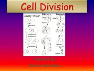
Mitosis + Cytokinesis = Cell division • Most cells in all eukaryotic organisms will divide many times throughout the life of the organism • Mitosis is the process by which a cell duplicates its genetic materials (chromosomes and prepares for cell division • Cytokinesis is the division of the rest of the cell into two different daughter cells • Prokaryotes also divide through binary fission, but this is NOT mitosis/cytokinesis • In animals, cell division occurs during embryonic development, growth, and wound healing • Errors during cell division can cause cell death or cancer
Mitosis reorganizes DNA in the cell • Prior to mitosis, the cell creates an exact duplicate of its DNA material • During mitosis, the two copies are reorganized, repackaged into two sets of chromosomes, and divided to opposite ends of the cell • In most organisms, mitosis is immediately followed by cytokinesis (the cell body dividing in two) • The original cell (mother cell) is identical to the two resulting cells (daughter cells)
Mitosis vocabulary • Nucleus – location of DNA inside the cell • Nuclear envelope – the membrane that separates the nucleus from the cytoplasm • DNA – an incredibly long molecule that contains genetic blue prints for cell behavior • Chromatin – a loosely bundled coil of DNA. Most of the time, DNA is organized in this form, “ a loose rope” • Histones – proteins which hold a DNA strand together in the form of chromatin • Chromosome – a highly organized form of chromatin, “a tightly wrapped and carefully knotted rope ” • Each chromosome is composed of two identical parts called chromatids • Chromatids: two halves of a chromosome which contain the same genetic information • DNA exists in the form of chromosomes only during mitosis • Each chromosome looks like an X • Centromere – a bundle of proteins which connects the two chromatids of a chromosome, the “knot at the center of the X“ • Microtubules – part of a cell’s cytoskeleton. These are tubes of protein which use to pull chromosomes apart and to opposite ends of a cell during mitosis. They are powered by ATP! • Mitotic spindle – How microtubules are organized during mitosis. This is a collection of microtubule fibers which is formed to coordinate the pulling of apart of chromosomes. • Metaphase plate – the line along which chromosomes are lined up during mitosis. This arrangement is coordinated by the mitotic spindle.
5 Phases of Mitosis • Mitosis is composed of five phases • Prophase • Prometaphase • Metaphase • Anaphase • Telophase • During each phase, different proteins in the cell perform specific roles to divide the two copies of genetic material • Cytokinesis occurs after mitosis to create two cells
Prophase • Chromatin is being organized into chromosomes inside the nucleus • Microtubules are organized into mitotic spindles in the cytoplasm • Nuclear envelope is dissolving • By the end of prophase • Chromosomes and mitotic spindle are fully organized • Nuclear envelope has disappeared
Prometaphase • Chromosomes move towards each other and into the center of the cell • Microtubules move into the nuclear region and begin to connect to chromosomes at the centromere • Microtubules organize into two mitotic spindles, one at each end of the cell
Metaphase • The mitotic spindle is fully organized, and has pulling the chromosomes to the center of the cell • The spindle aligns chromosomes so that each centromere is lined up along the metaphase plate (the center of the cell)
Anaphase • Each chromosome is pulled apart into two chromatids (halves) at the centromere • Microtubule fibers contract (using ATP!), pulling chromatids to opposite ends of the cell towards the two spindles
Telophase • Chromatids arrive at opposite ends of the cell and begin to unfold into loose coils of chromatin • New nuclear envelopes begin to form around the chromatin to create two nuclei • Spindle fibers disperse into the cytoplasm • The cell membrane begins to cleave in preparation for cytokinesis
Cytokinesis • The cell membrane is being pinched off to form two separate compartments • Cytoplasm and organelles are being divided between the two forming cells • At the end of cytokinesis, the membrane fuses to create two daughter cells which contain identical copies of DNA, and equal amounts of cytoplasm and organelles • Cell division is complete!
Cell division movies • http://www.youtube.com/watch?v=VlN7K1-9QB0 • http://www.youtube.com/watch?v=CzPGhYiGyZ8&feature=related • Embryonic division in the worm c.elegans: http://www.youtube.com/watch?v=zsgOl04PESI&NR=1 • http://www.cellsalive.com/mitosis.htm