Download
1 / 114
1.14k likes | 1.17k Views
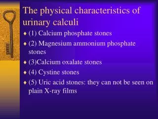
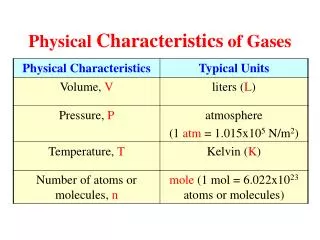
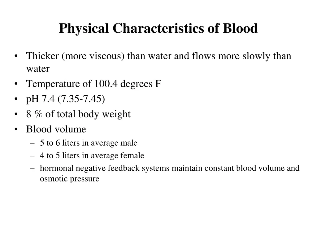
Physical Characteristics of Blood. Thicker (more viscous) than water and flows more slowly than water Temperature of 100.4 degrees F pH 7.4 (7.35-7.45) 8 % of total body weight Blood volume 5 to 6 liters in average male 4 to 5 liters in average female

E N D
Physical Characteristics of Blood • Thicker (more viscous) than water and flows more slowly than water • Temperature of 100.4 degrees F • pH 7.4 (7.35-7.45) • 8 % of total body weight • Blood volume • 5 to 6 liters in average male • 4 to 5 liters in average female • hormonal negative feedback systems maintain constant blood volume and osmotic pressure
Components of Blood • Hematocrit • 55% plasma • 45% cells • 99% RBCs • < 1% WBCs and platelets
Blood components • 55% plasma: 7 to 8% dissolved substances (sugars, amino acids, lipids & vitamins), ions, dissolved gases, hormones • ions are involved in membrane excitability, determination of fluid pH and osmotic pressure • most of the proteins in plasma are plasma proteins: provide a role in balancing osmotic pressure and water flow between the blood and extracellular fluid/tissues • loss of plasma proteins from blood – decreases osmotic pressure in blood and results in water flow out of blood into tissues – swelling • dispersed as colloid in the blood – opposite charge keeps them from “settling” out in blood • do not exit the blood due to their size – creates a protein gradient between blood and interstitial fluid • helps to determine interstitial and colloid pressures which determine bulk flow of fluid into tissues
Blood components • plasma proteins: albumin, globulins, clotting proteins (fibrinogen) • albumins – most abundant & common (~50% of plasma proteins) • contribute to most of the osmotic colloidal pressure • non-specifically bind many substances to help transport them through the blood • these substances are generally poorly soluble in water • e.g. bilirubin, bile salts, penicillin, steroid hormones
Blood components • plasma proteins: albumin, globulins, clotting proteins (fibrinogen) • globulins – 60 proteins divided into three classes (35% of plasma proteins) • alpha, beta and gamma • gamma globulins = immunoglobulins • alpha and beta globulins made by hepatocytes • e.g. lipoproteins, alpha-1-antitrypsin, transferrin, ferritin, C reactive protein • gamma globulins made by B cells • alpha and beta globulins bind substances for transport • e.g. thyroid hormone, iron, lipids, cholesterol • also involved in clotting • many inactive substances are alpha globulins • e.g. angiotensinogen (precursor to angiotensin)
Blood components • plasma proteins: albumin, globulins, clotting proteins (fibrinogen) • fibrinogen – cleaved by thrombin to produce a very stick mass of fibers made of fibrin • participate in clot formation • considered by some to be a beta-globulin
Blood: Cellular Elements • 45% of blood is the cellular elements or formed elements • produced from the Hematopoietic Stem Cells (HSCs) – found in the red bone marrow of the long bones and pelvis • form through a differentiation process called Hematopoiesis HSC
Blood: Cellular elements • 99% of blood cells are erythrocytes or RBCs • most numerous cell type in the body – 4 to 6 million per ul blood • flat, biconcave discs • provides a larger surface area for diffusion of oxygen across their membrane • thinness of the membrane allows rapid diffusion • very flexible membrane that allows their deformation for travel through thin capillaries
Erythrocytes • RBCs: • formed by differentiation of hematopoietic stem cells (HSCs) in the red bone marrow into myeloid stem cells erythrocytes • red bone marrow makes about 2 million RBCs per second! • first formed as an immature RBC = reticulocyte • still possess a nucleus and organelles • BUT lack mitochondria and cannot use the oxygen they transport for ATP synthesis • maturation of the reticulocyte causes loss of nucleus and organelles and the filling of the RBC with close to 250 million Hb molecules • RBCs have no nuclei or organelles but contain crucial enzymes to keep them alive for ~ 120 days • 1. glycolytic enzymes –for energy production (i.e. ATP) - RBCs rely on glycolysis and the enzymes of this pathway exclusively • 2. carbonic anhydrase– enzyme on RBC surface • plays role in dissolving of CO2 in blood plasma
Erythrocytes: Hemoglobin • pigment – naturally colored that is red due to its iron content • composed of a: • 1. globin portion • four, highly folded protein chains • 2 alpha chains & 2 beta chains • change their shapes as oxygen binds Hb
Erythrocytes: Hemoglobin • 2. heme component • one molecule of iron-based heme bound to each globin protein chain = 4 total • each heme can bind one oxygen – total binding capacity of 4 oxygen molecules per Hb • binding of the 1st O2 increases the binding efficiency of the other three O2 molecules • at sea level - Hb is 98% saturated with oxygen • saturation level can be affected by temperature (increase temp, decrease saturation) • saturation level can be affected by atmospheric pressure (decrease pressure, decrease saturation)
Hemoglobin • combines with • oxygen • hydrogen ions • carbon dioxide • the acidic portion of carbonic acid • carbon monoxide • occupies the oxygen binding sites • nitric oxide • binds to Hb in the lungs where it vasodilates pulmonary arterioles to ensure efficient transport of oxygenated blood from the lungs back to the heart
Erythropoiesis • process of making a RBC from a Hematopoeitic Stem Cell • HSC myeloid stem cell reticulocyte erythrocyte • RBCs produced first by the yolk sac of the embryo • then from myeloid stem cells in the red bone marrow • production is controlled at the level of the kidneys by the secretion of erythropoietin (EPO) • increases differentiation specifically of the myeloid stem cell RBC • increases release of mature RBCs or, if needed, the release of reticulocytes • synthetic EPO is now be made in the lab • used to boost RBC production during chemotherapy, diminishes the need for transfusions • role in blood-doping
Feedback Control of RBC Production • main stimulus for erythropoiesis = tissue hypoxia (cells not getting enough O2) • high altitude since air has less O2 • anemia • RBC production falls below RBC destruction • circulatory problems • Kidneys respond to hypoxia • release erythropoietin
RBC life-span and recycling • RBC lives only about 120 days • travels about 700 miles of vessels before destruction! • as the RBC ages the plasma membrane becomes very fragile and prone to rupture • destroyed by macrophages in the spleen, liver and bone marrow • major organ of destruction = spleen • small vessels tend to lyze the fragile RBCs as they travel through this organ • or internalized by macrophages • components of the RBC are recycled for other uses
RBC life-span and recycling • 1. macrophages of liver/spleen/bone marrow degrades the hemoglobin to its globin component and heme (2) • amino acid components of globin are reused for protein synthesis (3)
RBC life-span and recycling • 2. heme is degraded into free iron (Fe3+) and biliverdin • Fe3+ released into the blood (4) • Fe3+ is transported in blood attached to a protein called transferrin (5) • stored in liver, muscle or spleen (6) attached to a protein called ferritin • when needed – Fe3+ is sent to the bone marrow for hemoglobin synthesis (7&8)
RBC life-span and recycling • 9. in the liver - biliverdin is converted into bilirubin (11) - becomes part of bile • bile is secreted into the small intestine • the bilirubin is then into a series of compounds – end up expelled in urine as urobilin (13) or in the feces as stercobilin (14)
Hematocrit • Percentage of blood occupied by cells – since RBCs are 99% of these cells, hematocrit is a measurement of RBC count • female normal range • 38 - 46% (average of 42%) • male normal range • 40 - 54% (average of 46%) • testosterone
Polycythemia • too many RBCs (hematocrit over 65%) • dehydration, tissue hypoxia, blood doping in athletes • primary polycthemia • caused by a tumor-like condition of the bone marrow • overproduction of RBCs through increased differentiation of the myeloid stem cell • too many RBCs can increase the viscosity of the blood and result in dramatic decreases in blood pressure as frictional forces in the vessels increase – increases the workload of the heart • increased viscosity also slows the velocity of blood flow - reduce oxygen delivery to tissues • secondary polycthemia • appropriate EPO-induce adaptive mechanism to improve the blood’s oxygen carrying capacity • occurs at high altitudes or in people with chronic lung diseases
Anemia • Symptoms • oxygen-carrying capacity of blood is reduced • fatigue, cold intolerance & paleness • lack of O2 for ATP & heat production • Types of anemia • iron-deficiency = lack of absorption or loss of iron • type of nutritional anemia • failure to take in essential raw ingredients not made by the body • pernicious = lack of intrinsic factor for vitamin B12 absorption from the digestive tract • B12 is essential for normal RBC formation and maturation • binding of B12 to intrinsic factor allows its absorption • intrinsic factor – synthesized by the gastric mucosa • hemorrhagic = loss of RBCs due to bleeding (ulcer) • hemolytic = defects in cell membranes cause rupture • rupture of too many RBCs by external factors such as malaria (normal RBCs) or genetic disorders like sickle cell anemia (defective RBCs) • thalassemia = hereditary deficiency of hemoglobin • aplastic = destruction of bone marrow (radiation/toxins) • failure of the bone marrow to produce enough RBCs • may selectively destroy the ability to produce RBCs only • but may also destroy the myeloid stem cells – affect WBCs and platelets
Sickle-cell Anemia (SCA) • Genetic defect in hemoglobin molecule (Hb-S) that changes 2 amino acids in the globin protein • at low very O2 levels, RBC becomes deformed by changes in hemoglobin molecule within the RBC • sickle-shaped cells get stuck in capillary beds = causing occlusions and decreased blood flow to organs • also rupture easily = causing anemia & clots • Found among populations in malaria belt • Mediterranean Europe, sub-Saharan Africa & Asia • Person with only one sickle cell gene • increased resistance to malaria because RBC membranes leak K+ & lowered levels of K+ kill the parasite infecting the red blood cells
Blood Groups and Blood Types • RBC surfaces are marked by genetically determined glycoproteins & glycolipids • agglutinogens or isoantigens • distinguishes at least 24 different blood groups • ABO, Rh, Lewis, Kell, Kidd and Duffy systems
ABO Blood Groups • based on two glycolipid antigens on the surface of RBCs called A and B antigens • sugars attached to an RBC phospholipid • sugars are genetically determined • produced by specific enzymes = glycosyltransferases • what antigen you display tells you your blood type • the A and B antigens are connected to the RBC membrane through a “linker protein” called the H antigen – genetically determined
ABO Blood Groups • plasma also contains antibodies (or agglutinins) to the A or B antigens not found in your blood • blood type B – plasma has anti-A antibody that reacts with foreign antigen A
RH blood groups • Antigen was discovered in blood of Rhesus monkey • People with Rh antigens on RBC surface are Rh+ve. • Normal plasma contains no anti-Rh antibodies • Antibodies develop only in Rh-ve blood type & only with exposure to the antigen • e.g. accidental transfusion of positive blood • e.g. during a pregnancy with a positive blood type fetus • Transfusion reaction upon 2nd exposure to the antigen results in hemolysis of the RBCs in the donated blood
Hemolytic Disease of Newborn • Rh negative mom and Rh+ve fetus will have mixing of blood at birth • Mom’s immune system reacts to the foreign Rh antigen and makes antibodies • but also develops a memory to this foreign antigen and will make these antibodies the next time she is pregnant with an Rh+ve child • can cause hemolysis of developing fetal RBCs in the 2nd child • Mom's body will create Rh antibodies unless she receives a RhoGam shot • usually given as prenatal therapy to Rh negative mothers • RhoGam binds to loose fetal blood and removes it from body before she reacts • destroys the Rh antigen from the fetus before mother’s memory B cells develop
Thrombocytes: Platelets & clotting • Disc-shaped, 2 - 4 micron cell fragment with no nucleus • not whole cells! • do have organelles and cytosolic enzymes for generating energy from glucose • Normal platelet count is 150,000-400,000/drop of blood • Platelets form in bone marrow by following steps: • myeloid stem cells megakaryocyte-colony forming cells megakaryoblast megakaryocytes that fragment to form platelets • one megakaryocyte forms ~1000 platelets
Thrombocytes: Platelets & clotting • Short life span (5 to 9 days in bloodstream) • aged ones removed by macrophages in liver and spleen • numbers can be increased through the secretion of thrombopoietin (liver) which increases megakaryocyte development in the bone marrow and stimulates the formation of platelets • 30% of platelets are stored in the spleen and do not circulate • found in blood-filled spaces of the spleen = red pulp
Hemostasis • hemostasis = arrest of bleeding from a broken vessel • 3 steps: • 1) vascular spasm– constriction of smooth muscle layer in damaged vessel • intrinsic response triggered by physical damage • paracrine factors are released from the damaged endothelium • attract platelets to the area of damage • 2) platelet plug formation – aggregation of platelets to temporarily stop hemostasis and start the process of blot clot formation • 3) blood clotting – longer term hemostasis • remains until the damaged vessel wall is repaired
Platelet Plug Formation • plug formation requires the release of chemicals from activated platelets • a signal is required for their release = platelet adhesion • BUT platelets cannot stick to the smooth endothelium of intact vessels • BUT damage to the endothelial lining exposes collagen fibers to the platelet – results in their activation & formation of a platelet plug • the adhesion of platelets to collagen fibers is through an intermediary called vonWillebrand factor (vWF) • vWF is secreted by the damaged endothelium • vWF binds to the collagen fibers and becomes a “bridge” for the platelets to stick to • binding of the platelets results in their activation
Platelet Plug Formation • activated platelets store numerous paracrine factors in two kinds of cytoplasmic granules • 1. alpha granules • 4 platelet clotting factors = Pf1, Pf2, Pf3 and Pf4 • critical for initiating the next phase of hemostasis – clot formation • fibrinogen & clotting factor V – for clot formation • platelet-derived growth factor – for the repair of the damaged vessel
Platelet Plug Formation • 2. dense granules: • calcium – critical for the activity of clotting factors • thromboxane – chemotactic factor for other platelets • ADP - changes circulating platelet shape & causes them to become sticky – adhere to the first layer of aggregating platelets – results in the secretion of more ADP by the incoming platelets (positive feedback) • serotonin, epinephrine and thromboxane act as vasoconstrictors to reinforce the initial vascular spasm and attract more circulating platelets
Platelet Plug formation • platelet adhesion – by interaction with collagen fibers -results in their activation (2) platelet release of granule contents – by activated platelets (3) platelet aggregation – positive feedback system as aggregating platelets stimulate the arrival and activation of more platelets -plug becomes reinforced through the formation of sticky fibrin strands = BLOOD CLOT -aggregation is inhibited by the release of inhibiting factors by the adjacent normal endothelium – limits to plug to the area of damage
Blood Clotting • the blood clot stabilizes the weaker platelet plug and initiates healing of the damage vessel wall • ultimate step is the conversion of soluble fibrinogen (plasma protein) into insoluble & sticky fibrin by an enzyme called thrombin • substances required for clotting are: Ca+2, liver enzymes called clotting factors, fibrinogen and substances released by platelets & damaged tissues • thrombin– released by damaged cells, catalyzes the conversion of fibrinogen to fibrin • 12 liver clotting factors involved (CF VI is now known to be CF V)
The Clotting Cascade • clotting is a cascade of reactions in which each clotting factor activates the next in a fixed sequence resulting in the formation of fibrin threads • final steps: • 1. prothrombinase & Ca+2 convert prothrombin into thrombin • 2. thrombin convert fibrinogen into fibrin -final steps may be triggered through two possible paths 1. extrinsic pathway 2. intrinsic pathway
Common Pathway • requires formation of prothrombinase • comprised of activated clotting factors X (Xa) and V (Va) • Prothrombinase and Ca+2 catalyze the conversion of Prothrombinto Thrombin • Thrombin and Ca+2 convert Fibrinogen to Fibrin • Fibrin mesh traps blood cells = CLOT
Thrombin • converts soluble fibrinogen to insoluble fibrin threads • also activates fibrin stabilizing factor XIII • stabilizes the forming fibrin mesh • initiates several positive feedback effects • accelerates formation of Prothrombinase • activates factor V Va • also activates platelets to release factors which act to activate more factor X and therefore produces more thrombin (positive feedback) • can also act to increase the size of the clot by causing more platelet aggregation
Extrinsic Pathway • short-cut to clot formation • requires production of tissue factors produced externally from the blood vessel • start of the cascade is outside of the blood • damaged tissues produce and release Tissue Factor or thromboplastin into bloodstream – activated clotting factor VII • in the presence of Ca+2 - clotting factor X becomes activated and combines with activated clotting factor V to form prothrombinase • Prothrombinase forms in seconds
Intrinsic Pathway • everything required found is within the blood • drives clotting in damaged vessels and also induces clotting in blood samples in test tubes • activation of this pathways occurs either when: • endothelium is damaged & platelet plug forms • OR platelets themselves become damaged & release factors • requires several minutes for reaction to occur – occurs at the same time with platelet plug formation • also happens at the same time as the extrinsic pathway
Intrinsic Pathway • substances involved: Ca+2 and clotting factors XII, X and V • first factor formed – Factor XII (Hageman factor) • activated by contact with exposed collagen or glass surfaces • XIIa activates factor X • Xa combines with Va to form prothrombinase
12 Clotting: A summary Platelet aggregation
Clot Remodeling – The Fibrinolytic system • release of the fibrinolytic enzyme plasmin dissolves the clot • forms from plasminogen – inactive plasma protein present in the blood • plasminogen plasmin is stimulated by Factor XII • plasminogen becomes trapped in the forming clot, becomes activated to plasmin and slowly dissolves the clot as the tissue repairs itself • white blood cells then phagocytose the remaining parts of the clot as it dissolves • plasmin also functions to prevent clots from forming when not needed
Role of Vitamin K in Clotting • Normal clotting requires adequate vitamin K • fat soluble vitamin absorbed if lipids are present • absorption slowed if bile release is insufficient • Required for synthesis of 4 clotting factors by the hepatocytes • factors II (prothrombin), VII, IX and X • Produced by bacteria in large intestine • anti-coagulants called the coumarin drugs (heparin and warfarin) act by competing with vitamin K in the liver • inhibits the formation of the vitamin K-dependent clotting factors
Clotting Disorders: Hemophilia • Inherited deficiency of clotting factors • bleeding spontaneously or after minor trauma • subcutaneous & intramuscular hemorrhaging • nosebleeds, blood in urine, articular bleeding & pain • Hemophilia A - lacks factor VIII (males only) • most common • over 150 point mutations in the gene for factor VIII identified • factor VIII acts as a cofactor for the activation of factor X • Hemophilia B - lacks factor IX (males only) • less common • over 300 mutations in the DNA identified • Hemophilia C – lacks factor XI (males & females) • less severe because alternate clotting activators exist • Treatment is transfusions of fresh plasma or concentrates of the missing clotting factor
If clotting occurs in an unbroken vessel is called a thrombosis • clots can form in undamaged vessels if the body’s clotting and anti-clotting mechanisms are not kept balanced and in check • inappropriate clot attached to a vessel wall = thrombus • freely floating clot = embolus • thrombosis can result from several factors • 1. roughening of the endothelial lining during arterosclerotic plaque formation • 2. slow moving blood • 3. unbalanced fibrin-plasmin production • 4. widespread release of thromboplastin by tissues
Immunity • Immunity: ability of the body to defend itself from infectious agents, foreign cells, cancer cells • immune system has two functional divisions • innate immune system: non-specific immunity • divided into: cell-mediated and humoral (secreted) mediated • chemical and physical barriers • defensive chemicals: complement and inflammation • no memory • all forms of life • immune system recognizes some king of general molecular property marking the invader as foreign • usually a foreign carbohydrate or lipid in the cell wall • includes the response known as inflammation
Immunity • adaptive immune system: pathogen and antigen specific response • also divided into cell-mediated and humoral mediated • chemical defenses and lymphocytes • memory results • found only in jawed vertebrates
Cells of Immunity: WBCs WBCs can enter tissues where they function • cells of the lymphoid lineage • T and B lymphocytes (lymphocytes) • cells of the myeloid lineage • neutrophils, basophils, mast cells, eosinophils and monocytes, macrophages • BUT WBCs can also be classified based on appearance of the cytoplasm after staining • A. Granulocytes: neutrophils, macrophages, other phagocytes, eosinophils, basophils • B. Agranular: T and B cells