Download
1 / 19
200 likes | 330 Views
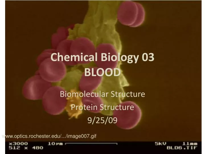
Chemical Biology 03 BLOOD. Biomolecular Structure Protein Structure 9/25/09. www.optics.rochester.edu/.../image007.gif . Chemical Biology 03 BLOOD. Biomolecular Structure Protein Structure Lecture 8: 9/25/09. Hierarchy of Protein Structure. Primary structure is determined by sequence

E N D
Chemical Biology 03BLOOD Biomolecular Structure Protein Structure 9/25/09 www.optics.rochester.edu/.../image007.gif
Chemical Biology 03BLOOD Biomolecular Structure Protein Structure Lecture 8: 9/25/09
Hierarchy of Protein Structure • Primary structure is determined by sequence • Secondary structure is α-helix, β-sheets, and turns or loops • Tertiary structure is packing of secondary structural elements • Quaternary is assembly of monomers into dimers, tetramers, etc. Fig 1.1 Branden & Tooz
Protein Structure "-HELIX • Favorable H-Bondingexists between the O of one amino acid residue and the N-H of a different amino acid residue four amino acids away. • A H bond is created when the electronegative lone pair electrons on the carbonyl oxygen cause the amide H to shift slightly away from N and towards O and form a weak bond. • O- - - H-N bond distance is ~2.9 D • bond energy is ~ 10 kJ/mole or less (compared with 400kJ for a C-H bond). • Must remember that these atoms would be interacting with solvent if they weren’t involved in this internal H bonding.
Protein Structure "-HELIX 13 atoms between hydrogen bonds. Xxxxxxxx
Protein Structure α-HELIX * In this highly idealized image of myoglobin, all of the helices are represented as colored ribbons, which trace their backbone. * For simplicity, all side groups are completely ignored. * How many helices do you see? See myoglobin file with Rasmol
Protein Structure α-HELIX • -R groups big and bulky side groups accommodated • charged amino acids project away from the cylinder • Ala, Glu, Leuand Metare good alpha helix formers • Pro, Gly, Tyrand Serare poor helix formers • Can see already from above observations that sequence determines structure. R groups on the outside of helix:
Protein Structure β strands and sheets • Anti parallel Beta Sheet: • Strands run in opposite N→C directions. • H bonds are made between different strands of the sheet, inter-strand H bonded network. • R groups: • alternate up and down the strand and across each strand, • are both up or both down across strands.
Protein Structure Beta Sheet • Beta sheet predicted (once again) by Pauling and found later experimentally in proteins. • Typical protein with extended beta sheet structure is fibroin, the major protein in silk which is a fabric produced by the silkworms of China and used for fabric since 2,000BC. Here, silk worms munch on mulberry leaves.. YUM
Protein Structure* Silk Note the H bonding within sheet that is inter-strand, and spacing pattern. Strand 1a Sheet A Strand 2a Strand 1b Sheet B Strand 2b
Protein Structure* Silk Note the R groups project above and below the sheets, and that the sheets aren’t perfectly flat, but are puckered in such a way that the knob of one R group fits into the upward pucker of sheet above. Intersheet van der Waals interactions govern polymer properties Sheet A Sheet B Identity of R groups determine tensile strengths, flexibilities, and price of different silks.
Protein Structure Beta Sheet • Parallel β sheet • Adjacent strands run in the same direction (-N to -C terminal) • H bonding pattern is evenly spaced Parallel Beta Sheet
Protein Structure Molecular Architecture • Beta twists: a common structural motif in which a mostly parallel beta sheet naturally twists. The twisted sheet is not purely parallel or antiparallel , but has a mixed orientation. Note from the N-term.: strand/loop/helix/loop strand/loop/helix/loop repeat Protein Thioredoxin
Protein StructureMolecular Architecture • Beta Barrels GFP, green fluorescent protein This beta stranded barrel and tight loops create a shielded lantern to hold the fluorescing group (cyclized triad of amino acids shown below). If group were exposed to solvent, the light would be extinguished.
Protein StructureLoops • Loops: Making the Connections • Loop regions connect regions of secondary structure, either helices or strands, or helices to strands. Previously thought to be unstructured random arrangements of amino acids, it is now realized that some regular features exist which can be described and in some cases rationalized. • They are usually at the surface of the molecule and therefore have polar charged or hydrophilic polar side groups. Glyis another favorite. Surprisingly, one can predict loop regions with more certainty than any other "structure". The carbonyl oxygen and amide proton hydrogen bond with the solvent.
Protein StructureLoops • Making Connections between α Helices • Alpha helices are often connected to each other by loop regions as seen in Mb. • The carbonyl and amide groups in the loops make hydrogen bonds with the solvent rather than internally. • Loop regions are often the sites of mutations, insertions, and deletions, since it seems you can perturb the loop and not affect the core structure. Myoglobin again, see loops
Protein Structure Loops LOOPS CONNECTING TWO ANTIPARALLEL BETA STRANDS:
Putting it together: What kinds of amino acid side groups would you expect to find on the surface of a protein like fibrinogen?