Download
1 / 37
370 likes | 495 Views
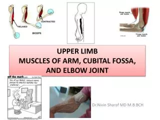
Appendicular &limb by dr.saro0ona. هذه المحاظرة ما هي إلا خدمه بسيطة لدفعتي الغالية 428 بأذن الله المحاظرة كاملة ,,وهي نسخه من الكتاب ,,وهدفي الأول منها إني أفيد ألي ما عندهم الكتاب أو ما يحبون الكتاب

E N D
هذه المحاظرة ما هي إلا خدمه بسيطة لدفعتي الغالية 428 • بأذن الله المحاظرة كاملة ,,وهي نسخه من الكتاب ,,وهدفي الأول منها إني أفيد ألي ما عندهم الكتاب أو ما يحبون الكتاب • و أبغى منكم دعوتين بظهر الغيب الأولى لوالدتي الغالية بالشفاء العاجل ,و الثانية لي بالتوفيق والتيسير بهالفاينل • ملاحظة: الي باللونالازرقهو من الكتاب ومو موجود بسلايداتدكتورتنا ولا دكتور العيال أختكم dr.saro0ona
Mesenchymal bones form in the limb buds duringthe(5) fifth week • mesenchymal bone models in the limbs undergo Chondrification to form hyaline cartilage bone models in the (6 )sixth week. • The clavicle initially develops by intramembranous ossification and it later forms growth cartilages at both ends • The bone models appear in a proximodistal sequence • Paterning in the developing limbs is regulated by homeobox containing (hox) genes
in the eighth week ,Ossification begins in the long bones & Initially occurs in the diaphysis from the primary ossification centers • By 12 weeks primary ossification centers have appeared in nearly all bones of the limbs. • The clavicles begin to ossify before any other bone in the body. • The femora are the next bones to show traces of ossification • First indication of ossification in cartilaginous model appear in the center of the future shaft, called primary center of ossification
Primary Ossification • Primary centers appear at different times in different bones .But ,Most of them develop between 7th and 12 weeks • Virtually all primary centers of ossification are present at birth • The part of the bone ossified from a primary center is the diaphysis
Secondary Ossification • Secondary ossification centers of the bones at knee are the first to appear in utero • The centers for the distal end of femur and proximal end of tibia appear during 34 to 38 weeks(last month) • Consequently they are present at birth • Most secondary centers of ossification appear after birth, • The part of bone ossified from the secondary centers called epiphysis
The bone forms from the primary center in the diaphysis do not fuse with that formed from the secondary centers in the epiphysis until the bone grows to its adult length • The delay enables lengthening of the bone to continue until the final size is reached
During bone growth, epiphysial plate intervenes between the diaphysis and epiphysis • The epiphysial plate is eventually replaced by bone development on each of its two sides, diaphysial and epiphysial • When this occurs, growth of the bone ceases
Limb Development • Limb development begins with the activation of a group of mesenchymal cells from the somatic layer of lateral mesoderm. • Limb buds first appear as elevations (swelling ) of the ventrolateral body wall toward the end of the 4th week ( 28 days). Homeobox- containing genes ( Hox ) regulate the limb development.
The limb buds appear as elevations of the ventrolateral body wall by end of 4th week • The limb buds form deep to a thick band of ectoderm • The upper limb buds are visible by 26 to 27 days • Lower limb buds appear 2 days later
Limb Bud • Limb buds elongate by the proliferation of the mesenchyme. • The upper limb buds appear disproportionately low on the embryo’s trunk because of the early development of the cranial half of the embryo. • The early stages of limb development are alike • for the upper and lower limbs • Because of their form & function, there are many distinct differences between the development of hand and foot
At the apex of each limb bud the ectoderm thickens to form and apical ectodermal ridge (AER) • The AER, a multilayered epitheleal structure , is induced by the underlying mesenchyme • Bone morphogenic protein signaling is required 4 its formation • AER exerts an inductive influence on the limb mesenchyme that initiates growth of limbs in proximal-distal axis
Digital Rays • By the end of 6th week, mesenchymal tissue in the hand plates has condensed to form digital rays • These mesenchymal condensations or finger buds outline the pattern of the digits • During the 7th week, similar condensations of mesenchyme form digital rays and toe buds in the foot plates • At the tip of each digital ray ,a part of AER induces development of the mesenchyme into the mesenchymalprimordia of the bones(phalanges) in the digits
The intervals between the digital rays are occupied by loose mesenchyme • Soon the intervening regions of mesenchyme break down forming notches between the digital rays • As the tissue breakdown progresses, separate digits are formed by the end of 8th week • Programmed cell death (apoptosis) is responsible for the tissue breakdown in the interdigital regions, and is probably mediated by bone morphogenetic proteins, signaling molecules of the transforming growth factor B (بيتا) • Blocking these cellular and molecular events could account for syndactyly, webbing or fusion of the fingers or toes
Final Stages of Limb Development • As the limb elongate, mesenchymal models of the bones are formed by cellular aggregation • Chondrification centers appear later in the 5th week. By the end of the 6th week, the entire limb skeleton is cartilaginous. • Osteogenesis of long bones begins in the 7th week from primary ossification centers in the middle of the cartilaginous models of long bones • ossification centers are present in all long bones by the 12th week • Ossification of the carpal (wrist) bones begins during the first year after birth
From the dermomyotome regions of the somites, myogenic precursor cells migrate into the limb bud and later differentiate into myoblasts ( precursors of muscle cells ). • As the long bones form, myoblasts aggregate and form a large muscle mass in each limb bud • In general this muscle mass separates into dorsal (extensor) and ventral (flexor) components • The mesenchyme in the limb bud gives rise to bones, ligaments, and blood vessels • The cervical and lumbosacralmyotome contribute to the muscles of the pectoral and pelvic girdles.
The developing upper limbs rotate in opposite directions and to different degrees • Rotations of Limbs: • The upper limbs rotate laterally through 90 degrees on their longitudinal axis • Now the future elbows point dorsally • Extensor muscles lie on the lateral and posterior aspects of the limb • The lower limbs rotate medially through 90 degrees • Now the future knees face ventrally • Extensor muscles lie on the anterior aspect of the lower limb
Developmentally, the radius and tibia are homologous bones • Ulna and fibula , just as the thumb and great toe are homologous digits • Synovial joints appear at the beginning of the fetal period , conceding functional differentiation of limb muscles and their innervations
Cutaneous Innervations of Limbs • There is strong relationship between the growth and rotation of the limb and the cutaneous segmental nerve supply of limbs. • Motor axons arising from the spinal cord enter the limb buds during the fifth week Grow into dorsal and ventral muscle masses. • Sensory axons enter the limb buds after the motor axons and use them for guidance. • Neural crest cells, the precursors of schwann cells, surround the motor and sensory nerve fibers in the limbs Form the neurolemmal and myelin sheaths.
During the 5th week, the peripheral nerves grow from the developing limb plexuses into mesenchyme of limb buds. • The spinal nerves are distributed in segmental bands, supplying both dorsal and ventral surfaces of the limb buds. • A dermatome is the area of skin supplied by a single spinal nerve and its ganglion, however , cutaneous nerve areas and dermatomes show considerable overlapping • As the limbs elongate, the cutaneous distribution of the spinal nerves migrates along the limbs and no longer reaches the surface in the distal part of the limbs
The original dermatomal pattern changes during growth of the limbs • An orderly sequence of distribution can still be recognized in the adult • In the upper limb, observe that the area supplied by C5 and C6 adjoin the areas supplied by T2, T1 and C8 but the overlap between them is minimal at the ventral axial line. • A cutaneous nerve area is the area of skin supplied by a peripheral nerve.
Because there is overlapping of dermatomes , a particular area of skin is not exclusively innervated by a single segmental nerve. • The limb dermatomes may be traced progressively down the lateral aspect of the upper limb and back up its medial aspect. • A comparable distribution of dermatosomes occur in the lower limb , which may be traced down the ventral aspect and then up the dorsal aspect of the lower limbs. • When the limbs descend they carry their nerves with them • This explains the oblique course of the nerves arising from the brachial and lumbosacral plexuses
Blood supply to limbs • The limb buds are supplied by branches of the intersegmental arteries which arise from the aorta and form a fine capillary network throughout the mesenchyme. • The primordial vascular pattern consists of a primary axial artery and its branches which drain into a peripheral marginal sinus. • Blood in the marginal sinus drain into a peripheral vein • The vascular pattern changes as the limbs develop, chievlygy angiogenesis (sprouting from existing vessels)
The new vessels coalesce with other sprouts to form new vessels • The primary axial artery becomes the brachial artery in the arm and the common interosseous artery in the forearm which has anterior and posterior interosseous branches. • The ulnar and radial arteries are terminal branches of the brachial artery. • As the digits form, the marginal sinus breaks up and final venous pattern is represented by basilic and cephalic veins and their tributaries develop.
In the thigh, the primary axial artery is represented by the deep artery of the thigh ( profundafemoris ). • In the leg, the primary axial artery is represented by the anterior and posterior tibial arteries.
anomalies of the limbs • ارجعوا لها من الكتاب ص372 و 373
تمت بحمد الله • لا تنسوني من صالح دعائكم